
Page 111 - TNFlipTest
P. 111
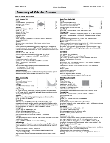
              Toronto Notes 2019 Valvular Heart Disease Summary of Valvular Disease
Cardiology and Cardiac Surgery C43
1
Auscultation: crescendo-decrescendo SEM radiating to R clavicle and carotid, musical
121
quality at apex (Gallavardin phenomenon), S4, soft S2 with paradoxical splitting, S3 (late)
Investigations
ECG: LVH and strain, LBBB, LAE, AFib
CXR: post-stenotic aortic root dilatation, calcifi
ed valve, LVH,
Echo: reduced valve area, pressure gradient, LVH, reduced LV function
Treatment
Asymptomatic: serial echos, avoid exertion S1 S2 Symptomatic: avoid nitrates/arterial dilators and ACEI in severe AS Surgery if: symptomatic or LV dysfunction
Surgical Options
Valve replacement: aortic rheumatic valve disease and trileaflet valve – prior to pregnancy (if AS significant)
121 selected patients who are not considered good candidates for surgery
Mitral Stenosis (MS) Etiology
Rheumatic disease most common cause, congenital (rare) Definition
– balloon valvuloplasty (in very young) S
Interventional Options 1
Percutaneous valve replacement (transfemorSal or transapicaSl approach) is an optioSn in
ECG: LVH, LAE SSS click
1
LAE, CHF 2
1
S1
CXR: LVH, LAE, aortic root dilatation
S OS 2
S 1
Echo/TTE: quantify AR, leaflet or aortic root anomalies
S1 S2 OS S1
Treatment
Asymptomatic: serial echos, afterload reduction (e.g. ACEI, nifedipine, hydralazine) Symptomatic: avoid exertion, treat CHF
Surgery if: NYHA class III-IV CHF; LV dilatation and/or LVEF <50% with/without symptoms
Surgical Options
Valve replacement: most patients
Valve repair: very limited role
Aortic root replacement (Bentall procedure): S
Cath: if >40 yr and surgical candidate – to assess for ischemic heart disease Exercise testing: hypotension with exercise
– when ascending aortic aneurysm present, valved conduit used
S1 S2os S1
 click
 Table 16. Valvular Heart Disease
Aortic Stenosis (AS)
Etiology
Congenital (bicuspid, unicuspid valve), calcification (wear and tear), rheumatic disease
Definition
Normal aortic valve area = 3-4 cm2 Mild AS >1.5 cm2
Moderate AS 1.0 to 1.5 cm2
Severe AS <1.0 cm2
Critical AS <0.5 cm2 Pathophysiology
S1
S 1
S 1
click
S2 os
S S
S1 S1
Aortic Regurgitation (AR)
Etiology
Supravalvular: aortic root disease (Marfan’s, atherosclerosis and dissecting aneurysm, connective tissue disease)
S Valvular: congenital (bicuspid aortic valve, S1 1 large VSD), IE
S2 S1
S2 S1
      S
1
S Symptoms 1 2 1
Exertional angina, syncope, dyspnea, PND, orthopnea, peripheral edema
Outflow obstruction → increased EDP → conce
ic LVH → LV failure → CHF, k
c subendocardial ischemia S S
S 2 1
Physical Exam
SSS
Physical Exam 1
Waterhammer pulse, bisferiens pulse, femoral-brachial sBP >20 (Hill’s test wide pulse pressure), hyperdynamic apex, displaced PMI, heaving apex
Auscultation: early decrescendo diastolic murmur at LLSB (cusp pathology) or RLSB (aortic root pathology), best heard sitting, leaSning forward, oSn fulol esxpiration, soft S1S,
absent S2, S3 (late)
Investigations
Narrow pulse pressure, brachial-radial delay, p
n
r
c
t
li
ulsus parvus et
tardus, sustained PMI 1
2
2
os
Acute Onset: IE, aortic dissection, trauma, failed prosthetic valve
Pathophysiology
Volume overload → LV dilatation → increased SV, high sBP and low dBP → increased wall tension → pressure overload → LVH (low dBP → decreased coronary perfusion) Symptoms
Usually only becomes symptomatic late in disease when LV failure develops Dyspnea, orthopnea, PND, syncope, angina S S
2
SSS 121
S S 121
      Severe MS is mitral valve area (MVA)
<1.5 cm2 S
Pathophysiology 1 2 1
MS → fixed CO and LAE → increased LA pressure → pulmonary vascular resistance
and CHF; worse with AFib (no atrial kick), tachycardia (decreased atrial emptying time) and pregnancy (increased preload)
Symptoms
SOB on exertion, orthopnea, fatigue, palpitations, peripheral edema, malar flush,
pinched and blue facies (severe MS)
Physical Exam
AFib, no “a” wave on JVP, left parasternal lift, palpable diastolic thrill at apex Auscultation: mid-diastolic rumble at apex, best heard with bell in left lateral decubitus position following exertion, loud S1, OS following loud P2 (heard best during expiration), long diastolic murmur, and short A2-OS interval correlate with worse MS
Investigations
ECG: NSR/AFib, LAE (P mitrale), RVH, RAD
CXR: LAE, CHF, mitral valve calcification
Echo/TTE: shows restricted opening of mitral valve
Cath: indicated in concurrent CAD if >40 yr (male) or >50 yr (female)
Treatment
Avoid exertion, fever (increased LA pressure), treat AFib and CHF, increase diastolic filling time (β-blockers, digitalis)
Surgery if: NYHA class III-IV CHF and failure of medical therapy
Invasive Options
Percutaneous balloon valvuloplasty: young rheumatic pts and good leaflet morphology (can be determined by echo), asymptomatic pts with moderate-severe MS, pulmonary HTN
Contraindication: left atrial thrombus, moderate MR
Open Mitral Commissurotomy: if mild calcification + leaflet/chordal thickening
– restenosis in 50% pts in 8 yr
Valve replacement: indicated in moderate-severe calcification and severely scarred leaflets
Mitral Regurgitation (MR)
Etiology
Mitral valve prolapse,
congenital cleft leaflets, LV
dilatation/aneurysm (CHF, DCM,
myocarditis), IE abscess, Marfan’s
syndrome, HOCM, acute MI, myxoma, S
mitral valve annulus calcification, 1
chordae/papillary muscle trauma/ischemia/rupture (acute), rheumatic disease Pathophysiology
Reduced CO → increased LV and LA pressure → LV and LA dilatation → CHF and pulmonary HTN
Symptoms
Dyspnea, PND, orthopnea, palpitations, peripheral edema
Physical Exam
S OS
S
S
S 2 1
Displaced hyperdynamic apex, left parasternal lift, apical thrill
S SOS S Auscultation: holosystolic murmur at apex, rad1iating to axilla ± m2 id-diastolic rumble1, loud
S2 (if pulmonary HTN), S3
Investigations
ECG: LAE, left atrial delay (bifid P waves), ± LVH CXR: LVH, LAE, pulmonary venous HTN
Echo: etiology and severity of MR, LV function, leaflets Swan-Ganz Catheter: prominent LA “v” wave Treatment
Asymptomatic: serial echos
Symptomatic: decrease preload (diuretics), decrease afterload (ACEI) for severe MR and poor surgical candidates; stabilize acute MR with vasodilators before surgery
Surgery if: acute MR with CHF, papillary muscle rupture, NYHA class III-IV CHF, AF, increasing LV size or worsening LV function, earlier surgery if valve repairable (>90% likelihood) and patient is low-risk for surgery
Surgical Options
Valve repair: >75% of pts with MR and myxomatous mitral valve prolapse – annuloplasty rings, leaflet repair, chordae transfers/shorten/replacement
Valve replacement: failure of repair, heavily calcified annulus
Advantage of repair: low rate of endocarditis, no anticoagulation, less chance of re- operation
  ©AnasNader2011 ©An©asANnaadserN2a0d1e1r2011 ©AnasNad©erA2n0a1s1Nader2011©AnasNad©erA20n1a1sNader2011©AnasNad©erA2n0a1s1Nader2011 ©AnasNader2011
© Anas Nader 2011 © Anas Nader 2011 © Anas Nader 2011 © Anas Nad©erA2n0a1s1Nader 2011 © Anas Nader 201©1 Anas Nader 2011 © Anas Nader 2011 © Anas Nader 2011 © Ana
