
Page 1130 - TNFlipTest
P. 1130
PL8 Plastic Surgery
Wounds
Toronto Notes 2019
•
• • •
• •
• • •
Wounds
wound:disruptionofthenormalanatomicalrelationshipsoftissueasaresultofinjury
Types of Wounds
laceration:cutortorntissue
abrasion:superficialskinlayerisremoved,variabledepth contusion:injurycausedbyforcefulblowtotheskinandsofttissue;entireouterlayerofskinintact,yet injured
avulsion:skinandsofttissue(generallyofalimb)forcefullyseparatedfromdeeperstructures, potentially compromising blood supply (i.e. “degloving”) or resulting in full detachment (amputation) puncturewounds:cutaneousopeningrelativelysmallascomparedwithdepth(e.g.needle),including bite wounds
crushinjuries:causedbycompression
burns: thermal, chemical, electrical
ulcers
Principles of Wound Healing
Table 7. Factors Influencing Wound Healing
Local (reversible/controllable)
Mechanical (local trauma, significant crush, avulsion, tension) Blood supply (ischemia/circulation)
Technique and suture materials
Retained foreign body
Infection
Venous HTN
Peripheral vascular disease Hematoma/seroma (h infection rate)
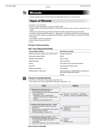
STAGES OF WOUND HEALING
General (often irreversible)
Age (affects healing rate)
Nutrition
Tobacco smoking
Alcohol consumption
Chronic illness (e.g. DM, cancer, CVD, renal failure) Immunosuppression (steroids, chemo)
Tissue irradiation
Genetic predisposition to abnormal healing (e.g. hypertrophic or keloid scarring, collagen vascular disease)
Skin type
• growthfactorsreleasedbytissuesplayanimportantrole
• scarismatureonceithascompletedthefinalstage,usuallyafter1-2yr
PHASE
1. Inflammatory Phase (Reactive) (Days 1-6)
• Limits damage, prevents further injury
• Debris and organisms cleared via inflammatory response:
• Neutrophils (24-48 h)
• Macrophages: critical to wound healing by
orchestrating growth factors for collagen production
(48-96 h)
• Lymphocytes: role poorly defined (5-7 d)
2. Proliferative Phase (Regenerative) (Day 4 – Week 3)
• Fibroblasts attracted and activated by macrophage growth factors
• Reparative process: re-epithelialization, matrix synthesis, angiogenesis (relieves ischemia)
• Tensile strength begins to increase at 4-5 d
3. Remodeling Phase (Maturation) (Week 3 – Year 1)
• Increasing collagen organization and stronger crosslinks • Type I collagen replaces Type III until normal 4:1
ratio achieved
• Peak tensile strength at 60 d – 80% of pre-injury strength
PROCESS
1. Hemostasis – vasoconstriction + platelet plug
2. Chemotaxis – migration of macrophages and PMN
1. Collagen synthesis (mainly type III) 2. Angiogenesis
3. Epithelialization
1. Contraction
2. Scarring
3. Remodeling of scar
Figure 15. Stages of wound healing
