
Page 1153 - TNFlipTest
P. 1153
Toronto Notes 2019 Craniofacial Injuries Craniofacial Injuries
• lowvelocityvs.highvelocityinjuriesdeterminedegreeofdamage
• fracturescausebruising,swelling,andtenderness→lossoffunction
• management:mostcanwait~5dforswellingtodecreasebeforeORIFrequired
Approach to Facial Injuries
• ATLSprotocol
• inspect,palpate,clinicalassessmentforinjurytounderlyingstructures(e.g.facialnerve,bonyinjuries,
septal hematoma, ocular involvement, etc.)
• tetanusprophylaxis
• radiologicalevaluation:CTscanwithfinecutsthroughtheorbit
• wound irrigation with NS/RL and remove foreign materials
• conservativedebridementofdetachedornonviabletissue
• repairatthetimeofpresentationwith4-0nylonsutureswhenthepatient’sgeneralconditionallows
• considerintracranialtrauma;ruleoutskullfracture
Investigations
• CT(goldstandard)
■ axial and coronal (specifically request 1.5 mm cuts): for fractures of upper and middle face, as well
as mandible
■ indicated for significant head trauma, suspected facial fractures, and pre-operative assessment
• panorexradiograph:showsentireupperandlowerjaw;bestforisolatedmandiblefracture,butpatient must be able to sit; however, if high clinical suspicion and negative panorex, CT should be done
Treatment Goals
• consultationwhenindicated(dentistry,ophthalmology)
• re-establishnormalocclusionifocclusionisanissue
• normaleyefunction(extraoculareyemovementsandvision) • restorestabilityoffaceandappearance
Mandibular Fractures
• twopointsofinjurysinceitisaringstructure(includesfracturesanddislocations) • commonlyatsitesofweakness(condylarneck,angleofmandible)
Etiology
• anteriorforce:bilateralfractures
• lateralforce:ipsilateralsubcondylarandcontralateralangleorbodyfracture • note:classifiedasopeniffractureintotoothbearingarea(alveolus)
Clinical Features
• pain,swelling,difficultyopeningmouth(“trismus”) • malocclusion,asymmetryofdentalarch
• damaged,loose,orlostteeth
• palpable“step”alongmandible
• numbnessinV3distribution
• intra-orallacerationsorhematoma(sublingual) • chindeviatingtowardsideofafracturedcondyle
Classification
Table 25. Mandibular Fracture Classifications by Anatomic Region
Plastic Surgery PL31
Patients with major facial injuries are at risk of developing upper airway obstruction (displaced blood clots, teeth, or fracture fragments; swelling of pharynx and larynx; loss of support of hyomandibular complex → retroposition of tongue); also at risk of ocular injury
Signs of Basal Skull Fracture
• Battle’s sign (bruised mastoid process) • Hemotympanum
• Raccoon eyes (periorbital bruising)
• CSF otorrhea/rhinorrhea
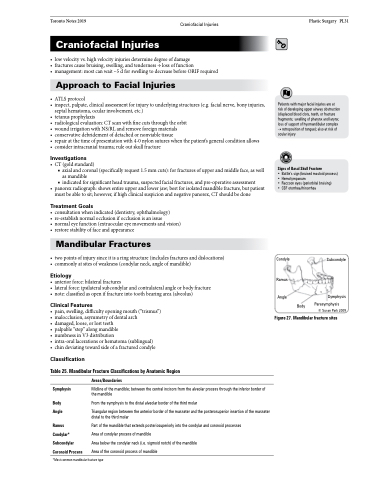
Condyle
Ramus Angle
Subcondyle
Symphysis Parasymphysis
© Susan Park 2009
Body
Figure27.Mandibularfracturesites
Symphysis
Body Angle
Ramus Condylar* Subcondylar
Coronoid Process
Areas/Boundaries
Midline of the mandible; between the central incisors from the alveolar process through the inferior border of the mandible
From the symphysis to the distal alveolar border of the third molar
Triangular region between the anterior border of the masseter and the posterosuperior insertion of the masseter distal to the third molar
Part of the mandible that extends posteriosuperiorly into the condylar and coronoid processes Area of condylar process of mandible
Area below the condylar neck (i.e. sigmoid notch) of the mandible
Area of the coronoid process of mandible
*Most common mandibular fracture type
