Page 1269 - TNFlipTest
P. 1269
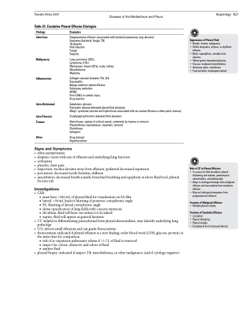
 Toronto Notes 2019 Diseases of the Mediastinum and Pleura Table 25. Exudative Pleural Effusion Etiologies
Respirology R23
Appearance of Pleural Fluid
• Bloody: trauma, malignancy
• White: empyema, chylous, or chyliform
effusion
• Black: aspergillosis, amoebic liver
abscess
• Yellow-green: rheumatoid pleurisy • Viscous: malignant mesothelioma • Ammonia odour: urinothorax
• Food particles: esophageal rupture
  Etiology
Infectious
Malignancy
Inflammatory
Intra-Abdominal
Intra-Thoracic Trauma
Other
Examples
Parapneumonic effusion (associated with bacterial pneumonia, lung abscess) Empyema (bacterial, fungal, TB)
TB pleuritis
Viral infection
Fungal Parasitic
Lung carcinoma (35%)
Lymphoma (10%)
Metastases: breast (25%), ovary, kidney Mesothelioma
Myeloma
Collagen vascular diseases: RA, SLE Pancreatitis
Benign asbestos related effusion Pulmonary embolism
ARMS
Post-CABG or cardiac injury Drug reaction
Subphrenic abscess
Pancreatic disease (elevated pleural fluid amylase)
Meigs’ syndrome (ascites and hydrothorax associated with an ovarian fibroma or other pelvic tumour)
Esophageal perforation (elevated fluid amylase)
Hemothorax: rupture of a blood vessel, commonly by trauma or tumours Pneumothorax (spontaneous, traumatic, tension)
Chylothorax
Iatrogenic
Drug-induced Hypothyroidism
   Signs and Symptoms
• oftenasymptomatic
• dyspnea:varieswithsizeofeffusionandunderlyinglungfunction
• orthopnea
• pleuriticchestpain
• inspection:tracheadeviatesawayfromeffusion,ipsilateraldecreasedexpansion
• percussion:decreasedtactilefremitus,dullness
• auscultation:decreasedbreathsounds,bronchialbreathingandegophonyatabovefluidlevel,pleural
friction rub
Investigations
• CXR
■ must have >200 mL of pleural fluid for visualization on PA film ■ lateral: >50 mL leads to blunting of posterior costophrenic angle ■ PA: blunting of lateral costophrenic angle
■ dense opacification of lung fields with concave meniscus
■ decubitus: fluid will layer out unless it is loculated
■ supine: fluid will appear as general haziness
• CT:helpfulindifferentiatingparenchymalfrompleuralabnormalities,mayidentifyunderlyinglung pathology
• U/S:detectssmalleffusionsandcanguidethoracentesis
• thoracentesis:indicatedifpleuraleffusionisanewfinding;orderbloodwork(LDH,glucose,protein)at
the same time for comparison
■ risk of re-expansion pulmonary edema if >1.5 L of fluid is removed ■ inspect for colour, character, and odour of fluid
■ analyze fluid
• pleuralbiopsy:indicatedifsuspectTB,mesothelioma,orothermalignancy(andifcytologynegative)
Role of CT in Pleural Effusion
• To assess for fluid loculation, pleural thickening and nodules, parenchymal abnormalities, and adenopathy
• Helps to distinguish benign from malignant effusion and transudative from exudative effusion
• May not distinguish empyema from parapneumonic effusion
Features of Malignant Effusion
• Multiple pleural nodules
Features of Exudative Effusion
• Loculation
• Pleural thickening
• Pleural nodules
• Extrapleural fat of increased density


