
Page 144 - TNFlipTest
P. 144
D10 Dermatology
Common Skin Lesions
Toronto Notes 2019
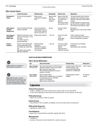
Table 8. Vascular Tumours
Hemangioma of Infancy
Spider Angioma (Campbell Telangiectasia)
Cherry Angioma (Campbell De Morgan Spot)
Pyogenic Granuloma
Clinical Presentation
Hot, firm red to blue plaques or tumours
Central red arteriole with slender branches, blanchable
Bright red to deep maroon, dome- shaped vascular papules, 1-5 mm Site: trunk
Less friable compared to pyogenic granulomas
Bright red, dome-shaped sessile or pedunculated friable nodule Sites: fingers, lips, mouth, trunk, toes
DDx: glomus tumour, nodular MM, SCC, nodular BCC
Pathophysiology
Benign vascular proliferation of endothelial lining
Can be associated with hyperestrogenic state (e.g. in liver disease, pregnancy, OCP) but more often is not
Benign vascular neoplasm
Rapidly developing hemangioma Proliferation of capillaries with erosion of epidermis and neutrophilia
Epidemiology
Appears shortly after birth; rarely may be congenital
Any age
>30 yr old
<30 yr old
Clinical Course
Appears shortly after birth, increases in size over months, then regresses
50% of lesions resolve spontaneously by 5 yr
Increase in number over time
Lesions do not fade in time
Lesions bleed infrequently
Lesion grows rapidly over wk-mo, then stabilizes
Lesion may persist indefinitely if untreated
Management
10% require treatment due to functional impairment (visual compromise, airway obstruction, high output cardiac failure) or cosmesis
Consider treatment if not gone by school age; topical timolol, propranolol; systemic corticosteroids; laser treatment; surgery
Reassurance
Electrodesiccation or laser surgery if patient wishes
Usually no treatment needed
Laser or electrocautery for small lesions Excision of large lesions if necessary
Surgical excision with histologic examination
Electrocautery; laser; cryotherapy
VASCULAR MALFORMATIONS
Table 9. Vascular Malformations
Pyogenic granuloma is a misnomer: it is neither pyogenic nor granulomatous
Venous Lake: benign blue or violaceous papular lesion occurring on the face, lips, and ears due to dilation of a venule. Distinguished from malignant pigmented lesions through diascopy, as compression blanches the lesion
Type
Nevus Flammeus
(Port-wine stain)
Nevus Simplex
(salmon patch)
Clinical Presentation
Red to blue macule present at birth that follows a dermatomal distribution, rarely crosses midline Most common site: nape of neck
Never spontaneously regresses but grows in proportion to the child
Pink-red irregular patches
Midline macule on glabella known as “Angel Kiss”; on nuchal region known as “Stork Bites”
Present in 1/3 of newborns
Majority regress spontaneously
Pathophysiology
Congenital vascular malformation of dermal capillaries; rarely associated with Sturge-Weber syndrome (V1, V2 distribution)
Congenital dilation of dermal capillaries
Management
Laser or make-up
No treatment required
Lipoma
Clinical Presentation
• singleormultiplenon-tendersubcutaneoustumoursthataresoftandmobile
• occursmostfrequentlyonthetrunk,andextremitiesbutcanbeanywhereonthebody
Pathophysiology
• adipocytesenclosedinafibrouscapsule
Epidemiology
• oftensolitaryorfewinnumber,ifmultiplecanbeassociatedwithraresyndromes
Differential Diagnosis
• angiolipoma, liposarcoma
Investigations
• biopsy only if atypical features (painful, rapid growth, firm)
Management
• reassurance
• excisionorliposuctiononlyifdesiredforcosmeticpurposes
