
Page 160 - TNFlipTest
P. 160
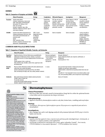
 D26 Dermatology DERMIS
Table 16. Comparison of Erysipelas and Cellulitis
Infections
Complications
Scarlet fever, streptococcal gangrene,
fat necrosis, coagulopathy Spreads via lymphatics
Uncommon
Toronto Notes 2019
    Erysipelas
Cellulitis
Clinical Presentation
Involves upper dermis
Confluent, erythematous, sharp raised edge, warm plaque, well demarcated Very painful (“St. Anthony’s fire”) Sites: face and legs
Systemic symptoms: fever, chills, headache, weakness (if present, sign of more serious infection)
Involves lower dermis/subcutaneous fat Unilateral erythematous flat lesion, often with vesicles poorly demarcated, not uniformly raised
Tender
Sites: commonly on legs
Systemic symptoms (uncommon): fever, leukocytosis, lymphadenopathy
Etiology
GAS
GAS, S. aureus (large sized wounds), H. influenzae (periorbital), Pasteurella multocida (dog/
cat bite)
Differential Diagnosis
DVT (less red, less hot, smoother), superficial phlebitis, contact dermatitis, photosensitivity reaction, stasis dermatitis, panniculitis, vasculitis
Same as erysipelas
Investigations
Clinical diagnosis: rarely do skin/blood culture
If suspect necrotizing fasciitis: do immediate biopsy and frozen section, histopathology
Same as erysipelas
Management
1st line: penicillin, cloxacillin or cefazolin 2nd line: clindamycin or cephalexin
If allergic to penicillin, use erythromycin
1st line: cloxacillin or cefazolin/cephalexin 2nd line: erythromycin or clindamycin Children: cefuroxime If DM (foot infections): TMP/SMX and metronidazole
 COMMON HAIR FOLLICLE INFECTIONS
Table 17. Comparison of Superficial Folliculitis, Furuncles, and Carbuncles
 Superficial Folliculitis
Furuncles (Boils)
Carbuncles
Antiseptic (Hibiclens®)
Topical antibacterial (fusidic acid, mupirocin, erythromycin, or clindamycin)
Oral cloxacillin for 7-10 d
Incise and drain large furuncles to relieve pressure and pain
If afebrile: hot wet packs, topical antibiotic
If febrile/cellulitis: culture blood and aspirate pustules (Gram stain and C&S)
Cloxacillin for 1-2 wk (especially for lesions near external auditory canal/ nose, with surrounding cellulitis, and not responsive to topical therapy)
Same as for furuncles
Clinical Presentation
Superficial infection of the hair follicle (versus pseudofolliculitis: inflammation of follicle due to friction, irritation, or occlusion)
Acute lesion consists of a dome-shaped pustule at the mouth of hair follicle
Pustule ruptures to form a small crust
Sites: primarily scalp, shoulders, anterior chest, upper back, other hair-bearing areas
Red, hot, tender, inflammatory nodules with central yellowish point, which forms over summit and ruptures
Involves subcutaneous tissue that arises from a hair follicle
Sites: hair-bearing skin (thigh, neck, face, axillae, perineum, buttocks)
Deep-seated abscess formed by multiple coalescing furuncles Usually in areas of thicker skin
Occasionally ulcerates
Lesions drain through multiple openings to the surface Systemic symptoms may be associated
Etiology
Normal non-pathogenic bacteria (Staphylococcus – most common; Pseudomonas – hot tub) Pityrosporum
S. aureus
S. aureus
Management
    Dermatophytoses
Clinical Presentation
          A Placebo-Controlled Trial of Antibiotics for Smaller Skin Abscesses
N Engl J Med 2017;376:2545-55
Purpose: To determine the appropriate management of uncomplicated skin abscesses.
• infectionofskin,hair,andnailscausedbydermatophytes(fungithatlivewithintheepidermalkeratin or hair follicle and do not penetrate into deeper structures)
Pathophysiology
• digestionofkeratinbydermatophytesresultsinscalyskin,brokenhairs,crumblingnails/onycholysis
Etiology
• Trichophyton,Microsporum,Epidermophytonspecies(Pityrosporumisasuperficialyeastandnota dermatophyte)
Investigations
• skinscrapings,hair,and/ornailclippingsanalyzedwithpotassiumhydroxide(KOH)preptolookfor hyphae and mycelia
Management
• topicalsasfirstlineagentsfortineacorporis/crurisandtineapedis(interdigitaltype):clotrimazole,or terbinafine or ciclopirox olamine cream applied bid
• oraltherapyisindicatedforonychomycosisortineacapitis:terbinafine(Lamisil®–livertoxicity, CYP2D6 inhibitor) or itraconazole (Sporanox® – CYP3A4 inhibitor, liver toxicity)
Study: Multi-center, prospective, double-blind trial involving outpatient adults and children with abscesses 5 cm or smaller, stratified by presence of surgically drainable abscess, abscess size, number of sites of skin infection, and presence of non-purulent cellulitis. Following incision and drainage, participants were randomly assigned to 10
d courses of clindamycin, TMP-SMX or placebo. Primary outcome was clinical cure 7 to 10 d after treatment end. Intention-to-treat analyses were conducted.
Results: Seven hundred and eighty-six participants
were enrolled (505 adults, 281 children). Ten days after therapy, the cure rate was similar between clindamycin and TMP-SMX (83.1% vs. 81.7%; p=0.73), and was higher than that of the placebo group (68.9%; p=0.001 for both comparisons). Among those who were cured, new infections at 1 month follow-up were less common in the clindamycin group than the TMP-SMX or placebo groups (6.8% vs. 11.1%; p=0.03 vs. 12.4%; p=0.06). Adverse events were more frequent with clindamycin than either of the other groups (21.9% s. 11.1% vs. 12.5%), though all resolved without sequelae.
Conclusions: Clindamycin or TMP-SMX in conjunction with incision and drainage for simple abscesses improves short-term outcomes compared to incision and drainage alone, though side-effects must be considered.
