
Page 196 - TNFlipTest
P. 196
ER16 Emergency Medicine
Traumatology
Toronto Notes 2019
Vascular Injuries
• realignlimb/applylongitudinaltractionandreassesspulses(e.g.Dopplerprobe) • surgicalconsult
• directpressureifexternalbleeding
Compartment Syndrome
• whentheintracompartmentalpressurewithinananatomicalarea(e.g.forearmorlowerleg)exceeds
Vascular injury/compartment syndrome is suggested by “The 6 Ps” Injury Compartment Syndrome - 6 Ps
Pulse discrepancies
Pallor
Paresthesia/hypoesthesia
Paralysis
Pain (especially when refractory to usual analgesics)
Polar (cold)
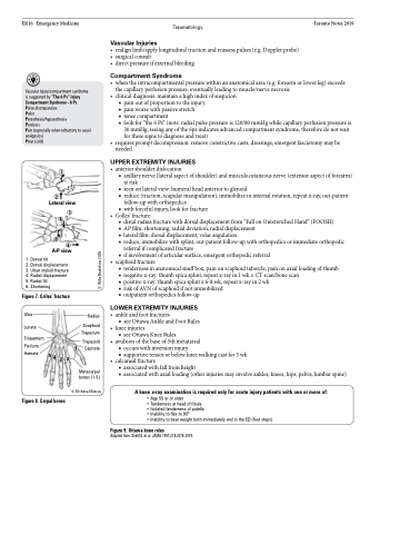
1
2
Lateral view
3 4
5
6
A-P view
1. Dorsal tilt
2. Dorsal displacement 3. Ulnar styloid fracture 4. Radial displacement 5. Radial tilt
6. Shortening
Figure 7. Colles’ fracture
Ulna Radius
Triquetrum
Pisiform Trapezoid
•
•
the capillary perfusion pressure, eventually leading to muscle/nerve necrosis clinical diagnosis: maintain a high index of suspicion
■ pain out of proportion to the injury
■ pain worse with passive stretch
■ tense compartment
■ look for “the 6 Ps” (note: radial pulse pressure is 120/80 mmHg while capillary perfusion pressure is
30 mmHg, seeing any of the 6ps indicates advanced compartment syndrome, therefore do not wait
for these signs to diagnose and treat) requirespromptdecompression:removeconstrictivecasts,dressings;emergentfasciotomymaybe needed
UPPER EXTREMITY INJURIES
• anteriorshoulderdislocation
■ axillary nerve (lateral aspect of shoulder) and musculocutaneous nerve (extensor aspect of forearm)
at risk
■ seen on lateral view: humeral head anterior to glenoid
■ reduce (traction, scapular manipulation), immobilize in internal rotation, repeat x-ray, out-patient
follow-up with orthopedics
■ with forceful injury, look for fracture
• Colles’ fracture
■ distal radius fracture with dorsal displacement from “Fall on Outstretched Hand” (FOOSH)
■ AP film: shortening, radial deviation, radial displacement
■ lateral film: dorsal displacement, volar angulation
■ reduce, immobilize with splint, out-patient follow-up with orthopedics or immediate orthopedic
referral if complicated fracture
■ if involvement of articular surface, emergent orthopedic referral
• scaphoidfracture
■ tenderness in anatomical snuff box, pain on scaphoid tubercle, pain on axial loading of thumb ■ negative x-ray: thumb spica splint, repeat x-ray in 1 wk ± CT scan/bone scan
■ positive x-ray: thumb spica splint x 6-8 wk, repeat x-ray in 2 wk
■ risk of AVN of scaphoid if not immobilized
■ outpatient orthopedics follow-up
LOWER EXTREMITY INJURIES
Lunate Trapezium
Scaphoid
• • •
ankle and foot fractures
■ see Ottawa Ankle and Foot Rules
knee injuries
■ see Ottawa Knee Rules
avulsion of the base of 5th metatarsal
■ occurs with inversion injury
■ supportive tensor or below knee walking cast for 3 wk
Hamate
Figure 8. Carpal bones
1 Capitate Metacarpal
bones (1-5)
© Elisheva Marcus
5 4 3 2
• calcanealfracture
■ associated with fall from height
■ associated with axial loading (other injuries may involve ankles, knees, hips, pelvis, lumbar spine)
A knee x-ray examination is required only for acute injury patients with one or more of:
• Age 55 yr or older
• Tenderness at head of fibula
• Isolated tenderness of patella
• Inability to flex to 90o
• Inability to bear weight both immediately and in the ED (four steps)
Figure 9. Ottawa knee rules
Adapted from: Stiell IG, et al. JAMA 1997;278:2075-2079.
© Willa Bradshaw 2005
