Page 225 - TNFlipTest
P. 225
 Toronto Notes 2019 Environmental Injuries Environmental Injuries
Heat Exhaustion and Heat Stroke
HEAT EXHAUSTION
• clinicalfeaturesrelatetolossofcirculatingvolumecausedbyexposuretoheatstress • “waterdepletion”:heatexhaustionoccursiflostfluidnotadequatelyreplaced
• “saltdepletion”:heatexhaustionoccurswhenlossesreplacedwithhypotonicfluid
HEAT STROKE
• life-threateningemergencyresultingfromfailureofnormalcompensatoryheat-sheddingmechanisms • dividedintoclassicalandexertionalsubtypes
• ifpatientdoesnotrespondrelativelyquicklytocoolingtreatments,considerotherpossibleetiologiesof
hyperpyrexia (e.g. meningitis, thyroid storm, anticholinergic poisoning, delirium tremens, infections), adverse drug events (including drug interactions)
Emergency Medicine ER45
Heat exhaustion may closely resemble heat stroke; heat exhaustion may eventually progress to heat stroke, so if diagnosis is uncertain treat as heat stroke
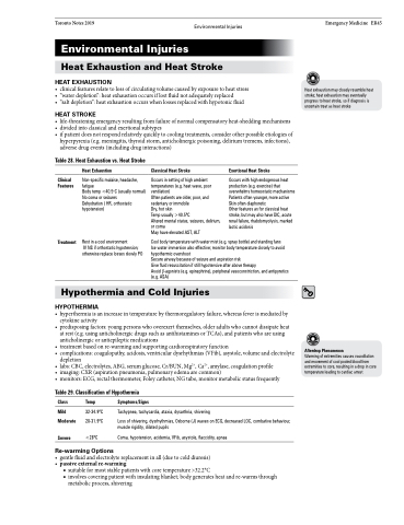
    Table 28. Heat Exhaustion vs. Heat Stroke
  Clinical Features
Treatment
Heat Exhaustion
Non-specific malaise, headache, fatigue
Body temp <40.5oC (usually normal) No coma or seizures
Dehydration ( HR, orthostatic hypotension)
Rest in a cool environment
IV NS if orthostatic hypotension;
otherwise replace losses slowly PO
Classical Heat Stroke
Occurs in setting of high ambient temperatures (e.g. heat wave, poor ventilation)
Often patients are older, poor, and sedentary or immobile
Dry, hot skin
Temp usually >40.5oC
Altered mental status, seizures, delirium, or coma
May have elevated AST, ALT
Exertional Heat Stroke
Occurs with high endogenous heat production (e.g. exercise) that overwhelms homeostatic mechanisms Patients often younger, more active Skin often diaphoretic
Other features as for classical heat stroke, but may also have DIC, acute renal failure, rhabdomyolysis, marked lactic acidosis
Cool body temperature with water mist (e.g. spray bottle) and standing fans
Ice water immersion also effective; monitor body temperature closely to avoid hypothermic overshoot
Secure airway because of seizure and aspiration risk
Give fluid resuscitation if still hypotensive after above therapy
Avoid β-agonists (e.g. epinephrine), peripheral vasoconstriction, and antipyretics (e.g. ASA)
   Hypothermia and Cold Injuries
HYPOTHERMIA
• hyperthermiaisanincreaseintemperaturebythermoregulatoryfailure,whereasfeverismediatedby cytokine activity
• predisposingfactors:youngpersonswhooverexertthemselves,olderadultswhocannotdissipateheat at rest (e.g. using anticholinergic drugs such as antihistamines or TCAs), and patients who are using anticholinergic or antiepileptic medications
• treatmentbasedonre-warmingandsupportingcardiorespiratoryfunction
• complications:coagulopathy,acidosis,ventriculardysrhythmias(VFib),asystole,volumeandelectrolyte
depletion
• labs: CBC, electrolytes, ABG, serum glucose, Cr/BUN, Mg2+, Ca2+, amylase, coagulation profile
• imaging: CXR (aspiration pneumonia, pulmonary edema are common)
• monitors:ECG,rectalthermometer,Foleycatheter,NGtube,monitormetabolicstatusfrequently
Table 29. Classification of Hypothermia
Afterdrop Phenomenon
Warming of extremities causes vasodilation and movement of cool pooled blood from extremities to core, resulting in a drop in core temperature leading to cardiac arrest
   Class
Mild Moderate
Severe
Temp
32-34.9oC 28-31.9oC
<28oC
Symptoms/Signs
Tachypnea, tachycardia, ataxia, dysarthria, shivering
Loss of shivering, dysrhythmias, Osborne (J) waves on ECG, decreased LOC, combative behaviour, muscle rigidity, dilated pupils
Coma, hypotension, acidemia, VFib, asystole, flaccidity, apnea
  Re-warming Options
• gentlefluidandelectrolytereplacementinall(duetocolddiuresis) • passiveexternalre-warming
■ suitable for most stable patients with core temperature >32.2°C
■ involves covering patient with insulating blanket; body generates heat and re-warms through
metabolic process, shivering


