
Page 426 - TNFlipTest
P. 426
GS24 General Surgery and Thoracic Surgery Small Bowel Obstruction Toronto Notes 2019
An acute abdomen + metabolic acidosis is bowel ischemia until proven otherwise
Investigations
• laboratory:leukocytosis(non-specific),andlacticacidosis(latefinding) ■ amylase, lactate, CK, and ALP can be used to observe progress
■ hypercoagulability workup if suspect venous thrombosis
• AXR:portalvenousgas,intestinalpneumatosis,andfreeairifperforation
• contrastCT:thickenedbowelwall,luminaldilatation,SMAorSMVthrombus,mesenteric/portal
venous gas, and pneumatosis
• CTangiographyisthegoldstandardforacutearterialischemia
Treatment
• fluidresuscitation,correctmetabolicacidosis,NPO,NGTdecompressionofstomach,andprophylactic broad-spectrum antibiotics; avoid vasoconstrictors and digitalis
• exploratorylaparotomytoassessextentofviability±segmentalresectionofnecroticintestine
■ if extent of bowel viability is uncertain, a second look laparotomy 12-24 h later is mandatory
• angiogram,embolectomy/thrombectomy,bypass/graft,mesentericendarterectomy,anticoagulation therapy, and percutaneous transluminal angioplasty ± stent
Carcinoid Syndrome Symptoms FDR
Flushing
Diarrhea
Right-sided heart failure
Tumours of Small Intestine
BENIGN TUMOURS
• 10xmorecommonthanmalignant
• usuallyasymptomaticuntillarge
• mostcommonsites:terminalileumandproximaljejunum • polyps
■ adenomas
■ hamartomas
■ FAP (see Familial Colon Cancer Syndromes, GS33) ■ juvenile polyps
• other:leiomyomas,lipomas,andhemangiomas
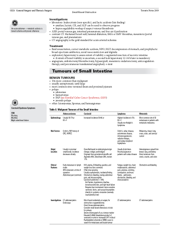
Table 8. Malignant Tumours of the Small Intestine
Epidemiology
Risk Factors
Origin/ Location
Clinical Features
Investigations
Adenocarcinoma
Usually 50-70 yr M>F
Crohn’s, FAP, history of CRC, HNPCC
Usually in proximal small bowel, incidence decreases distally
Early metastasis to lymph nodes
80% metastatic at time of operation
Abdominal pain (common)
CT abdomen/pelvis Endoscopy
Carcinoid
Increased incidence 50-60 yr
Classified based on embryological origin (foregut, midgut, and hindgut)
Originate from gut enterochromaffin cell Appendix 46%, distal ileum 28%, rectum 17%
N/V, anemia, GI bleeding, jaundice, and weight loss (less common)
Often slow-growing
Usually asymptomatic, incidental finding Obstruction, bleeding, crampy abdominal pain, and intussusception
Carcinoid syndrome (<10%)
Hot flashes, hypotension, diarrhea, bronchoconstriction, and right heart failure Requires liver involvement: lesion secretes serotonin, kinins, and vasoactive peptides directly to systemic circulation (normally inactivated by liver)
Most found incidentally at surgery for obstruction or appendectomy
Chest thorax/abdomen/pelvis
Consider small bowel enteroclysis to look for primary
Serum chromogranin A as a tumour marker Elevated 5-HIAA (breakdown product of serotonin) in urine or increased 5-HT in blood Radiolabelled octreotide or MIBG scans to search for metastases and locate tumour
Lymphoma
Highest incidence in 70s M>F
Usually non-Hodgkin’s lymphoma
Crohn’s, celiac disease, autoimmune disease, immunosuppression, radiation therapy,
and nodular lymphoid hyperplasia
Usually distal ileum Proximal jejunum in patients with celiac disease
Fatigue, weight loss, fever malabsorption, abdominal pain, anorexia, vomiting, constipation, and mass Rarely – perforation, obstruction, bleeding, and intussusception
CT abdomen/pelvis
Metastatic
Most common site of GI metastases in patients with metastatic melanoma
Melanoma, breast, lung, ovary, colon, and cervical cancer
Hematogenous spread from breast, lung, and kidney Direct extension from cervix, ovaries, and colon
Obstruction and bleeding
CT abdomen/pelvis
