
Page 429 - TNFlipTest
P. 429
Toronto Notes 2019
Appendix
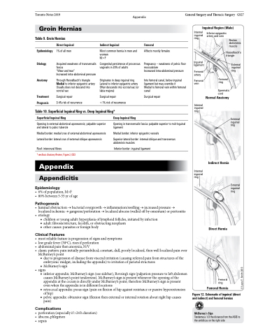
General Surgery and Thoracic Surgery GS27 Inguinal Region (Male)
Groin Hernias
Table 9. Groin Hernias
Internal inguinal ring
Inguinal ligament
Femoral artery
Femoral vein
Internal inguinal ring
Internal inguinal ring
Figure 12. Schematic of inguinal (direct and indirect) and femoral hernias
McBurney’sSign
Tenderness1/3thedistancefromtheASISto the umbilicus on the right side
Inferior epigastric artery and vein
Direct Inguinal
1% of all men
Acquired weakness of transversalis fascia
“Wear and tear”
Increased intra-abdominal pressure
Through Hesselbach’s triangle Medial to inferior epigastric artery Usually does not descend into scrotal sac
Surgical repair
3-4% risk of recurrence
Indirect Inguinal
Most common hernia in men and women
M>F
Congenital persistence of processus vaginalis in 20% of adults
Originates in deep inguinal ring Lateral to inferior epigastric artery Often descends into scrotal sac (or labia majora)
Surgical repair
<1% risk of recurrence
Femoral
Affects mostly females
Pregnancy – weakness of pelvic floor musculature
Increased intra-abdominal pressure
Into femoral canal, below inguinal ligament but may override it
Medial to femoral vein within femoral canal
Surgical repair
Rectus abdominis muscle
Epidemiology Etiology
Anatomy
Treatment Prognosis
Hesselbach’s triangle
External inguinal ring
External inguinal ring
Femoral ring
Spermatic cord
Normal Anatomy
Table 10. Superficial Inguinal Ring vs. Deep Inguinal Ring*
Superficial Inguinal Ring
Opening in external abdominal aponeurosis; palpable superior and lateral to pubic tubercle
Medial border: medial crus of external abdominal aponeurosis Lateral border: lateral crus of external oblique aponeurosis
Roof: intercrural fibres
*see Basic Anatomy Review, Figure 2, GS3
Appendix
Appendicitis
Epidemiology
• 6%ofpopulation,M>F
• 80%between5-35yrofage
Pathogenesis
Deep Inguinal Ring
Opening in transversalis fascia: palpable superior to mid-inguinal ligament
Medial border: inferior epigastric vessels
Superior-lateral border: internal oblique and transversus abdominis muscles
Inferior border: inguinal ligament
Indirect Hernia
External inguinal ring
• luminalobstruction→bacterialovergrowth→inflammation/swelling→increasedpressure→ localized ischemia → gangrene/perforation → localized abscess (walled off by omentum) or peritonitis
• etiology
■ children or young adult: hyperplasia of lymphoid follicles, initiated by infection ■ adult: fibrosis/stricture, fecolith, or obstructing neoplasm
■ other causes: parasites or foreign body
Clinical Features
• mostreliablefeatureisprogressionofsignsandsymptoms
• lowgradefever(38oC),risesifperforation
• abdominalpainthenanorexia,N/V
• classicpattern:paininitiallyperiumbilical;constant,dull,poorlylocalized,thenwelllocalizedpainover
McBurney’s point
■ due to progression of disease from visceral irritation (causing referred pain from structures of the
embryonic midgut, including the appendix) to irritation of parietal structures ■ McBurney’s sign
• signs
■ inferior appendix: McBurney’s sign (see sidebar), Rovsing’s sign (palpation pressure to left abdomen
causes McBurney’s point tenderness). McBurney’s sign is present whenever the opening of the appendix at the cecum is directly under McBurney’s point; therefore McBurney’s sign is present even when the appendix is in different locations
■ retrocecal appendix: psoas sign (pain on flexion of hip against resistance or passive hyperextension of hip)
■ pelvic appendix: obturator sign (flexion then external or internal rotation about right hip causes pain)
Complications
• perforation(especiallyif>24hduration) • abscess,phlegmon
• sepsis
Direct Hernia
Femoral ring
Femoral Hernia
Inguinal canal
© Laura E. Smith 2013
