
Page 570 - TNFlipTest
P. 570
H30 Hematology
Pathophysiology of TTP
• Large vWF multimers secreted by endothelial cells are typically rapidly cleaved by ADAMTS-13 protease
• Congenital TTP is due to a genetic deficiency in ADAMTS-13
• Antibodies against ADAMTS-13 are present in acquired TTP (the more common form)
Differential Diagnosis of TTP
• DIC
• HUS
• aHUS
• HELLP
• Catastrophic antiphospholipid Ab syndrome • Evans syndrome (AIHA + ITP)
Disorders of Primary Hemostasis Toronto Notes 2019 Thrombotic Thrombocytopenic Purpura and
Hemolytic Uremic Syndrome
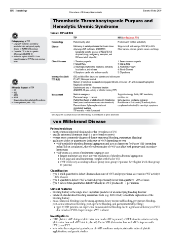
Table 24. TTP and HUS
Epidemiology Etiology
Clinical Features
Investigations (both TTP, HUS)
Management
TTP
Predominantly adult
Deficiency of metalloproteinase that breaks down ultra-large vWF multimers: ADAMTS13
Congenital (genetic absence of ADAMTS-13) Acquired (drugs, malignancy, transplant, and HIV-associated, idiopathic)
1. Thrombocytopenia
2. MAHA/TMA
3. Neurological symptoms: headache, confusion,
focal defects, and seizures
4. Symptoms can be mild and non-specific
HUS (see Pediatrics, P71)
Predominantly children and elderly
Shiga toxin (E. coli serotype O157:H7) in 90% Other bacteria, viruses, genetic causes, and drugs
1. Severe thrombocytopenia 2. MAHA/TMA
3. Acute kidney injury
4. Bloody Diarrhea
5. GI prodrome
CBC and blood film: decreased platelets and schistocytes
PT, aPTT, fibrinogen: normal
Markers of hemolysis: increased unconjugated bilirubin, increased LDH, and decreased haptoglobin Negative Coombs test
Creatinine and urea to follow renal function
ADAMSTS-13 gene, activity or inhibitor testing (TTP)
Medical emergency
Plasma exchange ± steroids
Platelet transfusion avoided unless life-threatening bleed (associated with microvascular thrombosis) Plasma infusion if plasmapheresis is not immediately available
TTP mortality ~90% if untreated
Supportive therapy (fluids, RBC transfusion, nutrition, etc.)
Some evidence for plasma exchange
Possible role of Eculizumab (C5 antibody blocks complement activation) for neurologic symptoms
Note: atypical HUS is a complex disease with different etiology, treatment depends on genetic abnormalities
von Willebrand Disease
Pathophysiology
• mostcommoninheritedbleedingdisorder(prevalenceof1%)
• usuallyautosomaldominant(type3isautosomalrecessive)
• womenmorecommonlydiagnosed(heavymenstrualbleeding,peripartumbleeding) • qualitativedefectorquantitativedeficiencyofvWFdependingontype
■ vWF needed for platelet adhesion/aggregation and acts as chaperone for Factor VIII (extending its half-life in circulation), therefore abnormality of vWF can affect both primary and secondary hemostasis
■ vWF exists as a series of multimers ranging in size
◆ largest multimers are most active in mediation of platelet adhesion/aggregation
◆ both large and small multimers complex with Factor VIII
◆ vWF levels vary according to blood group (non-group O patients have higher levels than group
O patients)
Classification
• type1:mildquantitativedefect(decreasedamountofvWFandproportionaldecreaseinvWFactivity) – 80% of cases
• type2:qualitativedefect(vWFactivitydisproportionallylowerthanquantity)–20%ofcases
• type3:severetotalquantitativedefect(virtuallynovWFproduced)–1permillion
Clinical Features
• bleedinghistoryisthesinglemostimportantpredictorofanunderlyingbleedingdisorder
• validated,standardizedbleedingassessmenttools(e.g.ISTH-BAT)tofacilitateexplorationofthe
bleeding history
• mucocutaneousbleeding(easybruising,epistaxis,heavymenstrualbleeding,peripartumbleeding,
post-dental extraction bleeding, post-operative bleeding, and gastrointestinal bleeding)
■ type 3 vWD patients can experience musculoskeletal bleeding due to significant deficiency in FVIII
due to lack of FVIII chaperoning as vWF is absent
Investigations
• CBC,platelet,vWF:Antigen(determinehowmuchvWFispresent),vWF:Ristocetincofactoractivity (determine how well vWF bind to platelet), Factor VIII (determine how well vWF chaperon with FVIII), and PTT
• teststofurthercategorizetype/subtypeofvWD:multimeranalysis,ristocetininducedplatelet agglutination, and genetic studies
