
Page 615 - TNFlipTest
P. 615
Toronto Notes 2019 Bone and Joint Infections
Clinical Presentation of Non-Gonoccocal Arthritis
• acuteonsetofpain,swelling,warmth,decreasedrangeofmotion±feverandchills;inchildren,refusal to weight bear
• mostofteninlargeweight-bearingjoints(knee,hip,ankle)andwrists
• usuallymonoarticular(polyarticularriskfactors:rheumatoidarthritis,endocarditis,GBS)
Investigations
• considerrheumatologiccausesformonoarthritis(seeRheumatology,RH3)
• gonococcal:bloodC&S,aswellasendocervical,urethral,rectal,andoropharyngealtesting
• non-gonococcal:bloodC&S
• arthrocentesis(synovialfluidanalysis)ismandatory:CBCanddifferential,Gramstain,C&S,examine
for crystals
■ infectious = opaque, increased WBCs (>15,000/mm3: likelihood of infection increases with
increasing WBCs), PMNs >90%, culture positive
■ growth of N. gonorrhoeae from synovial fluid is successful in <50% of cases
• ±plainx-ray:assessforosteomyelitis,providesbaselinetomonitortreatment
Treatment
• medical
■ empiric IV antibiotics: specific choice depends on clinical scenario and local guidelines; for most
adults, vancomycin + ceftriaxone is reasonable; for fully vaccinated children, cefazolin or cloxacillin
IV unless MRSA is a consideration – delay may result in joint destruction
■ Gram stain and cultures guide subsequent treatment
■ gonococcal: ceftriaxone + azithromycin, for concurrent treatment of C. trachomatis
■ non-gonococcal: antibiotics against Streptococcus spp. (2-3 wk IV f/b PO), S. aureus (4 wk IV
minimum), or GNB (4 wk)
• surgicalinterventionif(seeOrthopedics,OR11)
■ would consider surgical intervention on all cases of septic arthritis if possible ■ persistent positive joint cultures on repeat arthrocentesis
■ hip joint involvement, especially in pediatric population
■ prosthetic joint
• dailyjointaspirationsuntilculturesterile • physiotherapy
Prognosis
• gonococcal:respondswellafter24-48hofinitiatingantibiotics(usuallycompleterecovery)
• non-gonococcal:inchildren,generallygoodoutcomeiftreatedpromptly;inadults,upto50%
morbidity (decreased joint function/mobility)
Diabetic Foot Infections
Etiology
• neuropathy,peripheralvasculardisease,andhyperglycemiacontributetofootulcersthathealpoorly, and are predisposed to infection
• organismsinmildinfection:Streptococcusspp.,S.aureus
• organisms in moderate/severe infection: polymicrobial with aerobes (S. aureus, Streptococcus,
Enterococcus, GNB) and anaerobes (Peptostreptococcus, Bacteroides, Clostridium)
Clinical Presentation
• notallulcersareinfected
• considerinfectionif:probetobone(seebelow),ulcerpresent>30d,recurrentulcers,trauma,PVD,
prior amputation, loss of protective sensation, renal disease, or history of walking barefoot
• diagnosis of infected ulcer: ≥2 of the cardinal signs of inflammation (redness, warmth, swelling, pain)
OR the presence of pus
• ±crepitus,osteomyelitis,systemictoxicity
• visibleboneorprobetobone→osteomyelitis
• infection severity
■ mild = superficial (no bone/joint involvement)
■ moderate = deep (beneath superficial fascia, involving bone/joint) or erythema >2 cm
■ severe = infection in a patient with systemic toxicity (fever, tachypnea, leukocytosis, tachycardia,
hypotension)
Investigations
• curettagespecimenfromulcerbase,aspiratefromanabscessorbonebiopsy(resultsfromsuperficial swabs do not represent organisms responsible for deeper infection)
• bloodC&Siffebrile
• assessforosteomyelitisbyx-ray(althoughnotsensitiveinearlystages)orMRI/bonescanifhigh
clinical suspicion
■ ifinitialx-raynormal,repeat2-4wkafterinitiatingtreatmenttoincreasetestsensitivity
Infectious Diseases ID15
Intra-articular steroids are contraindicated until septic arthritis has been excluded
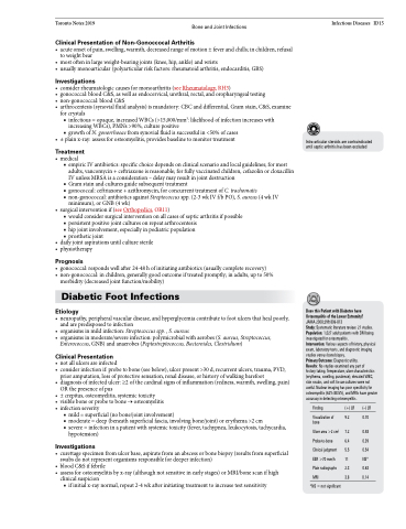
Does this Patient with Diabetes have Osteomyelitis of the Lower Extremity? JAMA 2008;299:806-813
Study: Systematic literature review. 21 studies. Population: 1,027 adult patients with DM being investigated for osteomyelitis.
Intervention: Various aspects of history, physical exam, laboratory tests, and diagnostic imaging studies versus bone biopsy.
Primary Outcome: Diagnostic utility.
Results: No studies examined any part of
history taking. Temperature, ulcer characteristics (erythema, swelling, purulence), elevated WBC, skin swabs, and soft tissue cultures were not useful. Nuclear imaging has poor specificity for osteomyelitis (62%-88.5%), and MRIs have greater accuracy in detecting osteomyelitis.
Finding
Visualization of bone
Ulcer area >2 cm2 Probe-to-bone Clinical judgment ESR >70 mm/h Plain radiographs MRI
*NS=notsignificant
(+) LR
9.2
7.2 6.4 5.5 11 2.3 3.8
(–) LR
0.70
0.48 0.39 0.54 NS* 0.63 0.14
