
Page 693 - TNFlipTest
P. 693
Toronto Notes 2019 Nuclear Medicine Medical Imaging MI25
Hyperparathyroidism
• mostcommoncauseisrenalfailure(secondaryhyperparathyroidism)
• chondrocalcinosisisacommoncomplication
• calciumcrystaldepositioninhyalinecartilageorfibrocartilage(includingarteriesandperi-articular
soft tissue)
• resorptionofbonetypicallyinhands(subperiostealandattufts),sacroiliacjoints(subchondral),skull
(“salt and pepper” appearance), subligamentous resorption (ischial tuberosity, trochanters and clavicle),
osteoclastoma (brown tumours)
• “ruggerjerseyspine”:band-likeosteosclerosisatsuperior/inferiormarginsofvertebralbodies
Paget’s Disease
• abnormalremodellinginvolvingsingleormultiplebones–especiallyskull,spine,pelvis • 3phases:1stphase=lytic,2ndphase=mixed(lytic/sclerotic),3rdphase=sclerotic
• coarseningofthetrabeculaewithboneexpansion
• bonesoftening/bowing
• bonescanwillrevealhighactivity,especiallyatboneends • thickenedcortex
• seeEndocrinology,E44
Nuclear Medicine
Brain
• 99mTc-exametazime(HMPAO)and99mTc-bicisate(ECD)imagingusedinSPECTtoassesscerebralblood flow and cellular metabolism, taken up predominantly in grey matter
■ used for dementia, traumatic brain injury, and to a lesser extent vasculitis, neuropsychiatric disorders, and occasionally stroke
■ most commonly used tracers to confirm brain death (i.e. absent blood flow to the brain and absent uptake on delayed planar and SPECT images in brain and brainstem, assuming study is technically adequate)
■ either tracer can be used for seizure imaging to assess for the most likely location of epileptogenic focus but usually must be made available for 24 h and the patient followed by a nurse who is competent to administer the activity at the time of seizure
• PETimagingassessesmetabolicactivitymostcommonlywith18FDG;usedfordementiaimaging,grade and stage of brain tumours, occasionally for seizure disorder imaging, and vasculitis; PET imaging with amyloid tracers for diagnosis of Alzheimer’s disease is becoming more common
• CSFimaging,intrathecaladministrationof111InDTPAtoevaluateCSFleakortodifferentiatenormal pressure hydrocephalus from brain atrophy
• CSFshuntevaluationforobstruction(mostcommonlyventriculoperitoneal)withsterileorpyrogen free 999mTc (usually) or 111In-DTPA; small quantity of activity is injected into the reservoir under sterile conditions and should flow freely into the peritoneal cavity by 45 min; maneuvers such as pumping the shunt, sitting the patient upright or ambulating are acceptable to encourage flow during this time
• adrenergicimagingoftheheartwithMIBGhasbeenusedtodifferentiatedementiaswithautonomic dysfunction (i.e. Lewy Body and Parkinson’s disease) from other forms of dementia (i.e. autonomic impairment associated with decreased MIBG activity in the heart)
Thyroid
Radioactive Iodine Uptake (see Endocrinology, E22)
• indexofthyroidfunction(trappingandorganificationofiodine)
• radioactive131IgivenPOtofastingpatient(smallquantity)
• measurepercentageofadministerediodinetakenupbythyroid
• increasedRAIU:toxicmultinodulargoitre,toxicadenoma,Graves’disease
• decreasedRAIU:subacutethyroiditis,lateHashimoto’sdisease,exogenousthyroidhormoneoriodine,
falsely decreased in patient with recent radiographic contrast studies, high dietary iodine (e.g. seaweed,
taking a “thyroid vitamin”)
• important–iodineuptakehelpsinthedifferentialofhyperthyroidismonly,nothypothyroidism
(exception is pediatrics)
Thyroid Imaging (Scintiscan)
• 99mTc-pertechnetateIVorradioactiveiodine(123I);mostCanadiansitesusepertechnetatetoreducecost • providesfunctionalanatomicdetail
• hot(hyperfunctioning)lesions:usuallybenign(e.g.adenoma,toxicmultinodulargoitre),cancer
unlikely (<1%) – No FNA
• cold(hypofunctioning)lesions:cancermustbeconsidereduntilbiopsynegativeeventhoughonly
6-10% are cancerous; decision to biopsy should be based on clinical and sonographic features
• isointensei.e.“warm”lesions:cancermustbeconsideredasanisointenselesionmayrepresentcold
nodules superimposed on normal tissue; if cyst suspected, correlate with U/S
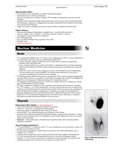
Figure 41. Multinodular goitre (top). Cold nodule (bottom)
