
Page 924 - TNFlipTest
P. 924
OP34 Ophthalmology
Ocular Manifestations of Systemic Disease
Toronto Notes 2019
Aflibercept, Bevacizumab, or Ranibizumab for Diabetic Macular Edema: 2 Year Result from a Comparative Effectiveness Randomized Clinical Trial
Ophthalmology 2016;123:1351-1359
All 3 anti-VEGF groups showed improvement of VA and deceased number of injections in year 2. Among eyes with worse baseline VA, aflibercept had superior 2 yr VA compared with bevacizumab, but superiority over ranibizumab in year 1 was no longer identified.
Effects of Medical Therapies on Retinopathy Progression in Type 2 DM
NEJM 2010;363:233-244
Purpose: To determine whether intensive glycemic control, combination therapy for dyslipidemia,
and intensive blood pressure control may limit the progression of DR in persons with type 2 DM. Methods: RCT with 10,251 participants with type 2 DM at high risk of cardiovascular disease. Intensive or standard treatment for glycemia (glycated hemoglobin level <6.0% or 7.0-7.9%), dyslipidemia (160 mg daily fenofibrate plus simvastatin or placebo plus simvastatin), or systolic blood- pressure control (target <120 or <140 mmHg). Results: Rates of progression of DR at 4 yr were 7.3% with intensive glycemia treatment vs. 10.4% with standard therapy (OR 0.67; 95% CI 0.51-0.87); 6.5% with fenofibrate for intensive dyslipidemia therapy vs. 10.2% with placebo (OR 0.60; 95% CI 0.42-0.87) and 10.4% with intensive blood pressure therapy vs. 8.8% with standard therapy (OR 1.23; 95% CI 0.84-1.79).
Conclusions: Intensive glycemic control and intensive combination treatment of dyslipidemia, but not intensive blood pressure control, reduced the rate of DR.
Extraocular Muscle Palsy
• usuallyCNIIIinfarct
• pupilusuallysparedindiabeticCNIIIpalsy,butptosisisobserved • mayinvolveCNIVandVI
• usually recover within few months
Optic Neuropathy
• visualacuitylossduetoinfarctionofopticdisc/nerve
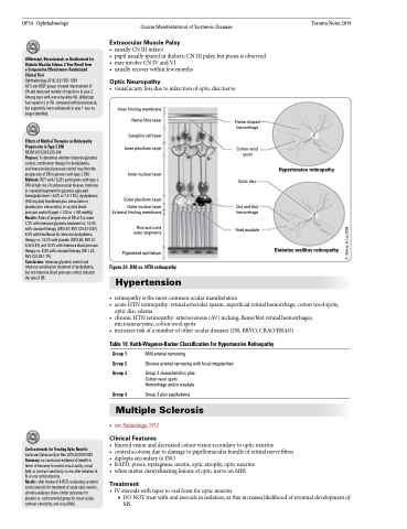
Inner limiting membrane Nerve fibre layer
Ganglion cell layer Inner plexiform layer
Inner nuclear layer
Outer plexiform layer
Outer nuclear layer External limiting membrane
Rod and cone outer segments
Pigmented epithelium
Figure 24. DM vs. HTN retinopathy
Hypertension
Flame shaped hemorrhage
Cotton wool spots
Optic disc
Dot and blot hemorrhage
Hard exudate
Hypertension retinopathy
Diabetes mellitus retinopathy
• retinopathyisthemostcommonocularmanifestation
• acuteHTNretinopathy:retinalarteriolarspasm,superficialretinalhemorrhage,cottonwoolspots,
optic disc edema
• chronicHTNretinopathy:arteriovenous(AV)nicking,flame/blotretinalhemorrhages,
microaneurysms, cotton wool spots
• increasesriskofanumberofotheroculardiseases(DR,BRVO,CRAO/BRAO)
Table 10. Keith-Wagener-Barker Classification for Hypertensive Retinopathy
Group 1 Group 2 Group 3
Group 4
Mild arterial narrowing
Obvious arterial narrowing with focal irregularities
Group 2 characteristics plus: Cotton wool spots Hemorrhage and/or exudate
Group 3 plus papilledema
Corticosteroids for Treating Optic Neuritis
Cochrane Database Syst Rev 2015;8:CD001430 Summary: no conclusive evidence of benefit in terms of recovery to normal visual acuity, visual field, or contrast sensitivity six mo after initiation of IV or oral corticosteroids.
Results: after review of 6 RCTs evaluating systemic corticosteroids for treatment of acute optic neuritis, all meta-analyses show similar outcomes for placebo vs. corticosteroid group for visual acuity, contrast sensitivity, and visual field.
Multiple Sclerosis
• seeNeurology,N52
Clinical Features
• blurredvisionanddecreasedcolourvisionsecondarytoopticneuritis
• centralscotomaduetodamagetopapillomacularbundleofretinalnervefibres • diplopiasecondarytoINO
• RAPD, ptosis, nystagmus, uveitis, optic atrophy, optic neuritis
• white matter demyelinating lesions of optic nerve on MRI
Treatment
• IVsteroidswithtapertooralformforopticneuritis
■ DO NOT treat with oral steroids in isolation, as this increases likelihood of eventual development of
MS
© Sherry H. Lai 2006
