
Page 1134 - TNFlipTest
P. 1134
PL12 Plastic Surgery
Wounds
Toronto Notes 2019
Table 9. Recommended Dressings for Wound Type
Wound Depth
Superficial Deep
Exudate Level
Lightly exuding
Any exudate level
Light to moderately exuding wounds
Moderately to heavily exuding wounds
Dressing Material
Films (Opsite®), hydrogels (Intrasite®, Nu-gel®, Duoderm®) Contact layers
Amorphous gels, hydrogels, hydrocolloids (Duoderm®, Tegaderm®), collagen, hypertonic saline gauze (Mesalt®)
Foams (Mepilex®, Allevyn®), alginates (Sorbsan®, Kalto-stat®), hypertonic saline gauze, hydrofibre (Aquacel®)
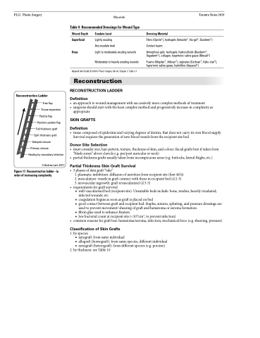
Reconstruction Ladder
Free flap
Tissue expansion
Pedicle flap Random pattern flap
Full thickness graft Split thickness graft
Delayed closure Primary closure
Healing by secondary intention ©Andrea Lam 2017
Figure 17. Reconstructive ladder - in order of increasing complexity
Adapted from Grabb & Smith’s Plastic Surgery, 6th ed. Chapter 3, Table 3.3
Reconstruction
RECONSTRUCTION LADDER
Definition
• anapproachtowoundmanagementwithsuccessivelymorecomplexmethodsoftreatment
• surgeonsshouldstartwiththeleastcomplexmethodandprogressivelyincreaseincomplexityas
appropriate
SKIN GRAFTS
Definition
• tissuecomposedofepidermisandvaryingdegreesofdermis,thatdoesnotcarryitsownbloodsupply. Survival requires the generation of new blood vessels from the recipient site bed
Donor Site Selection
• mustconsidersize,hairpattern,texture,thicknessofskin,andcolour(facialgraftsbestiftakenfrom “blush zones” above clavicle e.g. pre/post auricular or neck)
• partialthicknessgraftsusuallytakenfrominconspicuousareas(e.g.buttocks,lateralthighs,etc.)
Partial Thickness Skin Graft Survival
• 3phasesofskingraft“take”
1. plasmatic imbibition: diffusion of nutrition from recipient site (first 48 h) 2. inosculation: vessels in graft connect with those in recipient bed (d 2-3) 3. neovascular ingrowth: graft revascularized (d 3-5)
• requirementsforgraftsurvival
■ well-vascularized bed (recipient site). Unsuitable beds include: bone, tendon, heavily irradiated,
infected wounds, etc.
■ coagulation begins as soon as graft is placed on bed
■ good contact between graft and recipient bed. Staples, sutures, splinting, and pressure dressings are
used to prevent movement/ shearing of graft and hematoma or seroma formation
■ fibrin glue used to enhance fixation
■ low bacterial count at recipient site (<105/cm3, to prevent infection)
• commonreasonsforgraftloss:hematoma/seroma,infection,mechanicalforce(e.g.shearing,pressure)
Classification of Skin Grafts
1. by species
■ autograft: from same individual
■ allograft (homograft): from same species, different individual ■ xenograft (heterograft): from different species (e.g. porcine)
2. by thickness: see Table 10
