Page 1138 - TNFlipTest
P. 1138
 PL16 Plastic Surgery
Ulcers Toronto Notes 2019
CLINICAL FEATURES
• painoutofproportiontoclinicalfindingsandbeyondborderoferythema
• edema,tenderness,±crepitus(subcutaneousgasfromanaerobes),±flu-likesymptoms(e.g.nausea,
fever, diarrhea, dizziness, malaise)
• overlyingskinchangesincludingblisteringandecchymoses
• patientsmaylookdeceptivelywellatfirst,butmayrapidlybecomeverysick/toxic
• latefindings
■ skin turns dusky blue and black (secondary to thrombosis and necrosis) ■ induration, formation of bullae
■ cutaneous gangrene, subcutaneous emphysema
Investigations
• aclinicaldiagnosis
• CTscanonlyifsuspectitisnotnecrotizingfasciitis(lookingforabscess,gascollection,myonecrosis
and possible source of infection)
• severelyelevatedCK:usuallymeansmyonecrosis(latesign)
• bedsideincision,exploration,andincisionalbiopsywhenrulingoutconditions,clinicalpresentationis
not supportive, or difficult exam
• duringincisionalbiopsy,oftensee“dishwaterpus”(GroupAinfection)andahemostateasilypassed
along fascial plane (fascial biopsy to rule out in equivocal situations)
Treatment
• vigorousresuscitation(ABCs)
• urgentsurgicaldebridement:removeallnecrotictissue,copiousirrigationwithplansforrepeatsurgery
in 24-48 h
• IVantibiotics:asappropriateforclinicalscenario;considerpenicillin4millionIUIVq4hand/or
clindamycin 900 mg IV q6h until final cultures available (the combination can be synergistic if Group A
strep)
• urgentconsultationwithinfectiousdiseasespecialistisrecommended
Ulcers
Lower Limb Ulcers
Traumatic Ulcers (Acute)
• failureofwoundtoheal,usuallyduetocompromisedbloodsupplyandunstablescar,secondaryto pressure or bacterial colonization/infection
• usuallyoverbonyprominence±edema±pigmentationchanges±pain
• treatment:involvementofvascularsurgery.Anydebridementofulcerandcompromisedtissuesmust
be preceded by ABIs and vascular Doppler. Ulcers or compromised tissues left to heal via secondary intention with dressings may need reconstruction with local or distant flap in select cases, vascular status of limb must be assessed clinically and via vascular studies (i.e. ABI, duplex Doppler)
     Non-Traumatic Ulcers (Chronic)
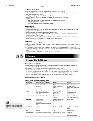
Table 14. Venous vs. Arterial vs. Diabetic Ulcers
    ABI in diabetics can be falsely normal due to incompressible arteries secondary to plaques/ calcification
Characteristic
Cause
History
Common Distribution Appearance
Wound Margins
Depth Surrounding Skin
Venous (70% of vascular ulcers)
Valvular incompetence Venous HTN
Dependent edema, trauma Rapid onset ± thrombophlebitis, varicosities
Medial malleolus (“Gaiter” locations)
Yellow exudates
Granulation tissue
Varicose veins
Brown discolouration of surrounding skin
Irregular
Superficial
Venous stasis discolouration (brown)
Arterial
2° to small and/or large vessel disease (be aware of risk factors)
Arteriosclerosis, claudication Usually >45 yr
Slow progression
Distal locations (e.g. lower limb, feet)
Pale/white, necrotic base ± dry eschar covering
Even (“punched out”)
Deep
Thin, shiny, dry skin; hairless, cool
Diabetic
Peripheral neuropathy: decreased sensation Atherosclerosis: microvascular disease
DM
Peripheral neuropathy Trauma/pressure
Pressure point distribution (more likely metatarsal heads)
Necrotic base
Irregular or “punched out” or deep
Superficial/deep
Thin, dry skin ± hyperkeratotic border Hypersensitive/ischemic


