
Page 1143 - TNFlipTest
P. 1143
Toronto Notes 2019 Burns Burn Wound Healing
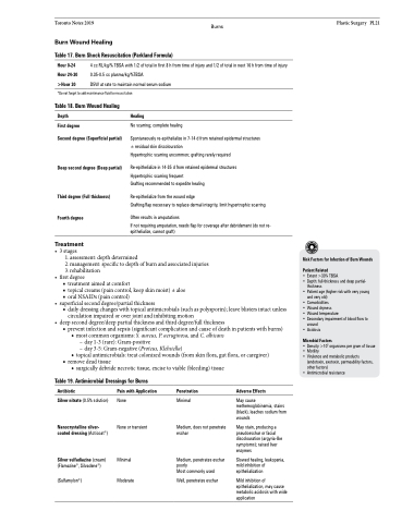
Table 17. Burn Shock Resuscitation (Parkland Formula)
4 cc RL/kg/% TBSA with 1/2 of total in first 8 h from time of injury and 1/2 of total in next 16 h from time of injury 0.35-0.5 cc plasma/kg/%TBSA
D5W at rate to maintain normal serum sodium
Plastic Surgery PL21
Hour 0-24
Hour 24-30
>Hour 30
*Do not forget to add maintenance fluid to resuscitation
Table 18. Burn Wound Healing Depth
First degree
Second degree (Superficial partial)
Deep second degree (Deep partial)
Third degree (Full thickness) Fourth degree
Treatment
Healing
No scarring; complete healing
Spontaneously re-epithelialize in 7-14 d from retained epidermal structures ± residual skin discolouration
Hypertrophic scarring uncommon; grafting rarely required
Re-epithelialize in 14-35 d from retained epidermal structures Hypertrophic scarring frequent
Grafting recommended to expedite healing
Re-epithelialize from the wound edge
Grafting/flap necessary to replace dermal integrity, limit hypertrophic scarring
Often results in amputations
If not requiring amputation, needs flap for coverage after debridement (do not re- epithelialize, cannot graft)
• 3stages
1. assessment: depth determined
2. management: specific to depth of burn and associated injuries 3. rehabilitation
• firstdegree
■ treatment aimed at comfort
■ topical creams (pain control, keep skin moist) ± aloe ■ oral NSAIDs (pain control)
• superficialseconddegree/partialthickness
■ daily dressing changes with topical antimicrobials (such as polysporin); leave blisters intact unless
circulation impaired or over joint and inhibiting motion
• deepseconddegree/deeppartialthicknessandthirddegree/fullthickness
■ prevent infection and sepsis (significant complication and cause of death in patients with burns) ◆ most common organisms: S. aureus, P. aeruginosa, and C. albicans
– day 1-3 (rare): Gram-positive
– day 3-5: Gram-negative (Proteus, Klebsiella)
◆ topical antimicrobials: treat colonized wounds (from skin flora, gut flora, or caregiver)
Risk Factors for Infection of Burn Wounds
Patient Related
• Extent >30% TBSA
• Depth: full-thickness and deep partial-
thickness
• Patient age (higher risk with very young
and very old)
• Comorbidities
• Wound dryness
• Wound temperature
• Secondary impairment of blood flow to
wound • Acidosis
Microbial Factors
• Density >105 organisms per gram of tissue
• Motility
• Virulence and metabolic products
(endotoxin, exotoxin, permeability factors,
other factors)
• Antimicrobial resistance
■ remove dead tissue
◆ surgically debride necrotic tissue, excise to viable (bleeding) tissue
Table 19. Antimicrobial Dressings for Burns
Antibiotic
Silver nitrate (0.5% solution)
Nanocrystalline silver- coated dressing (Acticoat®)
Silver sulfadiazine (cream) (Flamazine®, Silvadene®)
(Sulfamylon®)
Pain with Application
None
None or transient
Minimal Moderate
Penetration
Minimal
Medium, does not penetrate eschar
Medium, penetrates eschar poorly
Most commonly used
Well, penetrates eschar
Adverse Effects
May cause methemoglobinemia, stains (black), leaches sodium from wounds
May stain, producing a pseudoeschar or facial discolouration (argyria-like symptoms); raised liver enzymes
Slowed healing, leukopenia, mild inhibition of epithelialization
Mild inhibition of epithelialization, may cause metabolic acidosis with wide application
