
Page 1265 - TNFlipTest
P. 1265
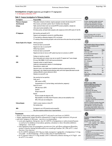
 Toronto Notes 2019 Pulmonary Vascular Disease Investigations (if highly suspicious, go straight to CT angiogram)
• seeEmergencyMedicine,ER33
Table 21. Common Investigations for Pulmonary Embolism
Respirology R19
D-dimer is elevated in patients with recent surgery, cancer, inflammation, infection, and severe renal dysfunction. It has good sensitivity and negative predictive value, but poor specificity and positive predictive value
Classic ECG finding of PE is S1-Q3-T3 (inverted T3), but most commonly see only sinus tachycardia
Clinical Prediction Rule for Pulmonary Embolism
J Thromb Hemost 2000;83:416-420
Wells’ Criteria
Risk Factors Points
Clinical signs of DVT 3.0 No more likely alternative diagnosis 3.0 (using H&P, CXR, ECG)
Immobilization or surgery in previous 4 wk 1.5 Previous PE/DVT 1.5 HR >100 beats/min 1.5 Hemoptysis 1.0 Malignancy 1.0
Clinical Probability
Low (0-2) 3% Intermediate (3-6) 28% High (>6) 78% Modified Wells’: >4 PE likely; ≤4 PE unlikely
JAMA 2006
PE Rule Out Criteria (PERC)
Prospective Multicentre Evaluation of the Pulmonary Embolism Rule Out Criteria
J Thromb Hemost 2008;6:772
• Agelessthan50yr
• Heartratelessthan100bpm
• Oxyhemoglobinsaturation≥95percent
• No hemoptysis
• Noestrogenuse
• NopriorDVTorPE
• Nounilaterallegswelling
• No surgery or trauma requiring hospitalization
within the past 4 wk
Acute PE can probably be excluded without further diagnostic testing if the patient meets all PERC criteria AND there is a low clinical suspicion for PE, according to either the Wells’ criteria or a low gestalt probability determined by the clinician prior to diagnostic testing for PE.
Evaluation of a Suspected Pulmonary Embolism
Low clinical probability of embolism D-dimer (+ve) → CT scan (+ve) → ruled in
(–ve)→ruledout (–ve)→ruledout Intermediate or high probability
CT scan (–ve) → ruled out
(+ve) → ruled in Notes
• Use D-dimers only if low clinical probability, otherwise, go straight to CT
• IfusingV/Qscans(CTcontrastallergyorrenalfailure): • NegativeV/Qscanrulesoutthediagnosis
• High probability V/Q scan only rules in the diagnosis if
have high clinical suspicion
• InconclusiveV/QscanrequireslegU/StolookforDVTor
    Investigation
Pulmonary Angiogram (Gold Standard) D-Dimer
CT Angiogram
Venous Duplex U/S or Doppler
ECG CXR
V/Q Scan
Purpose/Utility
Filling defect indicative of embolus; negative angiogram excludes clinically relevant PE More invasive, and harder to perform than CT, therefore done infrequently
Highly sensitive D-dimer result can exclude DVT/PE if pretest probability is already low Little value if pretest probability is high
If D-dimer positive, will need further evaluation with compression U/S (for DVT) and/or CT (for PE)
Both sensitive and specific for PE
Diagnosis and management uncertain for small filling defects
CT may identify an alternative diagnosis if PE is not present
CT scanning of the proximal leg and pelvic veins can be done at the same time and may be helpful
With leg symptoms
Positive test rules in proximal DVT
Negative test rules out proximal DVT
Without leg symptoms
Positive test rules in proximal DVT
Negative test does not rule out a DVT: patient may have non-occlusive or calf DVT
Findings not sensitive or specific
Sinus tachycardia most common; may see non-specific ST segment and T wave changes RV strain, RAD, RBBB, S1-Q3-T3 with massive embolization
Frequently normal; no specific features
Atelectasis (subsegmental), elevation of a hemidiaphragm
Pleural effusion: usually small
Hampton’s hump: cone-shaped area of peripheral opacification representing infarction
Westermark’s sign: dilated proximal pulmonary artery with distal oligemia/decreased vascular markings (difficult to assess without prior films)
Dilatation of proximal PA: rare
Very sensitive but low specificity Order scan if:
CXR normal, no COPD
Contraindication to CT (contrast allergy, renal dysfunction, pregnancy) Avoid V/Q scan if:
CXR abnormal or COPD Inpatient
Suspect massive PE
Results:
Normal: excludes the diagnosis of PE
High probability: most likely means PE present, unless pre-test probability is low 60% of V/Q scans are nondiagnostic
Useful to assess massive or chronic PE Not routinely done
No diagnostic use in PE (insensitive and nonspecific) May show respiratory alkalosis (due to hyperventilation)
                            Echocardiogram ABG
Treatment
   • admitforobservation(stablepatientswithDVTonlymaybesenthomeonLMWH)
• oxygen:supplementaloxygenshouldbeadministeredtotargetanoxygensaturation≥90percent • painrelief:analgesicsifchestpain–narcoticsoracetaminophen
■ acute anticoagulation: therapeutic-dose SC LMWH or fondaparinux or unfractionated heparin or oral factor Xa inhibitors (rivaroxaban, apixaban, edoxaban) or direct thrombin inhibitors (dabigatran) – start ASAP
■ anticoagulation stops clot propagation, prevents new clots, and allows endogenous fibrinolytic system to dissolve existing thromboemboli over months get baseline CBC, INR, aPTT ± renal function ± liver function
■ for SC LMWH: dalteparin 200 U/kg once daily, enoxaparin 1 mg/kg bid, or fondaparinux 5-10 mg once daily – no lab monitoring – avoid or reduce dose in renal dysfunction
■ for IV heparin: bolus of 75 U/kg (usually 5,000 U) followed by infusion starting at 20 U/kg/h – aim for aPTT 2-3x control
CT
