
Page 1277 - TNFlipTest
P. 1277
Toronto Notes 2019
Neoplasms
Respirology R31
Investigations
• CXR:alwayscomparewithpreviousCXR
• CTdensitometryandcontrastenhancedCTofthorax
• sputumcytology:usuallypooryield
• biopsy(bronchoscopicorpercutaneous)orexcision(thoracoscopyorthoracotomy):ifclinicaland
radiographic features do not help distinguish between benign or malignant lesion
■ if at risk for lung cancer, biopsy may be performed regardless of radiographic features
■ if a biopsy is non-diagnostic, whether to observe, re-biopsy, or resect will depend on the level of
Pulmonary neoplasms may present as
a solitary pulmonary nodule identified incidentally on a radiographic study (~10% of cases) or as symptomatic disease (most cases)
Adenocarcinoma present in a non-smoker may be due to endothelial growth factor receptor mutation
Corona Radiata Sign on Chest CT
• Fine striations that extend linearly from a nodule in a spiculated fashion
• Highly associated with malignancy
Carcinoids
• Early onset (40-60 yr)
• Most are central and can produce
symptoms and signs of bronchial
obstruction
• Hemoptysis is present in ~50% of cases
suspicion
• watchfulwaiting:repeatCXRand/orCTscanat3,6,12mo
• PETscancanhelpdistinguishbenignfrommalignantnodules
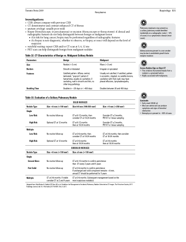
Table 32. CT Characteristics of Benign vs. Malignant Solitary Nodule
Parameters
Size Borders Features
Doubling Time
Benign
Nodule (<3 cm) Smooth or lobulated
Calcified pattern: diffuse, central, laminated, “popcorn” pattern if hamartoma, usually no cavitation; if cavitating, wall is smooth and thin, no other lung pathology
Doubles in <20 days or >400 days
Malignant
Mass (>3 cm)
Irregular or spiculated
Usually not calcified; if calcified, pattern is eccentric, stippled, no satellite lesions, cavitation with thick wall, may have pleural effusions, lymphadenopathy
Doubles between 20 and 400 days
Size >8 mm (>250 mm2)
Consider CT a 3 months, PET/CT or tissue sampling
Consider CT a 3 months, PET/CT or tissue sampling
CT at 3-6 months, then consider CT at 18-24 months
CT at 3-6 months then at 18-24 months
persistence years
Table 33. Evaluation of a Solitary Pulmonary Nodule
Nodule Type
Single
Low Risk
High Risk
Multiple Low Risk
High Risk
Nodule Type
Single
Ground Glass
Part Solid
Multiple
Size <6 mm (<100 mm2)
No routine follow-up Optional CT at 12 months
No routine follow-up Optional CT at 12 months
Size <6 mm (<100 mm2)
No routine follow-up No routine follow-up
CT at 3-6 months. If stable consider CT at 2 and 4 years
SOLID NODULES
Size 6-8 mm (100-250 mm2)
CT at 6-12 months, then consider CT at 18-24 months
CT at 6-12 months then at 18-24 months
CT at 3-6 months, then consider CT at 18-24 months
CT at 3-6 months then at 18-24 months
SUBSOLID NODULES
Size ≥6 mm (>100 mm2)
CT at 6-12 months to confirm then CT every 2 years until 5
CT at 3-6 months to confirm persistence
If unchanged and solid component remains <6 mm, annual CT should be performed for 5 years
CT at 3-6 months. Subsequent management based on the most suspicious nodule(s)
Adapted from: MacMahon H, Naidich DP, Goo JM, et al. Guidelines for Management of Incidental Pulmonary Nodules Detected on CT Images. The Fleischner Society 2017. Radiology Journal. doi:10.1148/radiol.2017161659. Feb 23 2017.
