Page 1279 - TNFlipTest
P. 1279
 Toronto Notes 2019 Introduction to Intensive Care Introduction to Intensive Care
Intensive Care Unit Basics
• goalistoprovidestabilizationforcriticallyillpatients:hemodynamic,respiratory,orcardiacinstability, or need for close monitoring
Lines and Catheters
• arteriallines
■ monitor beat-to-beat blood pressure variations, obtain blood for routine ABGs ■ common sites are the radial and femoral arteries
• centralvenouscatheter(centralline)
■ administer IV fluids, monitor CVP, insert pulmonary artery catheters
■ administer TPN and agents too irritating for peripheral line (e.g. vasopressors, chemotherapy) ■ common sites: internal jugular vein, subclavian vein, femoral vein
• pulmonaryarterialcatheter
■ balloon guides the catheter from a major vein to the right heart
■ measurespulmonarycapillarywedgepressure(PCWP)viaacatheterwedgedindistalpulmonaryartery ■ PCWP reflects the LA and LV diastolic pressure (barring pulmonary venous or mitral valve disease) ■ indications (now used infrequently due to associated complications)
◆ diagnosis of shock states, primary pulmonary HTN, valvular disease, intracardiac shunts, cardiac tamponade, PE
◆ assessment of hemodynamic response to therapies
◆ differentiation of high- versus low-pressure pulmonary edema
◆ management of complicated MI, multiorgan system failure and/or severe burns, or
hemodynamic instability after cardiac surgery ■ absolute contraindications
◆ tricuspid or pulmonary valve mechanical prosthesis ◆ right heart mass (thrombus or tumour)
◆ tricuspid or pulmonary valve endocarditis
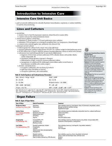
Table 34. Useful Equations and Cardiopulmonary Parameters
Respirology R33
               BSA = [Ht (cm) + Wt (kg) – 60]/100 SV = CO / HR
CI = CO / BSA
SVRI = [(MAP – RAP) 80]/CI
P:F ratio = PaO2 / FiO2
PCWP = LVEDP
SVI = CI / HR
RV Ejection Fraction = SV / RVEDV
PP = sBP – dBP
MAP = 1/3 sBP + 2/3 dBP = dBP + 1/3 PP
Intensive versus Conventional Glucose Control in Critically Ill Patients
NEJM 2009: 360;1283-1297
Purpose: To assess whether intensive glucose control improves mortality in critically ill patients. Study: Prospective, randomized controlled trial. Population: 6,104 patients expected to require ICU treatment for 3 or more consecutive days. Intervention: Patients were randomized to insulin therapy regimens with intensive (blood glucose 4.5-6 mM) or conventional (blood glucose 10 mM or less) glucose control targets. Intravenous insulin therapy was used to maintain blood glucose in target range.
Primary Outcome: Death from any cause within 90 days after randomization.
Results: The odds ratio for death in the intensive control group was 1.14 (95% CI 1.02-1.28; P=0.02) and this effect did not differ between surgical and medical patients. Severe hypoglycemia (blood glucose <2.2 mM) was significantly more common in the intensive management group (6.8% versus 0.5%; P<0.001).
Conclusion: Intensive insulin therapy in ICU patients increased mortality compared to blood glucose targeting of less than 10 mM with a number needed to harm of 38.
  BSA = body surface area; CI = cardiac index; CO = cardiac output; dBP = diastolic blood pressure; HR = heart rate; LVEDP = left ventricular end diastolic pressure; MAP = mean arterial pressure; PCWP = pulmonary capillary wedge pressure; PP = pulse pressure; RAP = right atrial pressure; RVEDV = right ventricular end diastolic volume; sBP = systolic blood pressure; SV = stroke volume; SVI = stroke volume index; SVRI = systemic vascular resistance index
 Organ Failure
Table 35. Types of Organ Failure
  Type of Failure
Respiratory Failure
(see Respiratory Failure, R26) Cardiac Failure
(see Cardiology and Cardiac Surgery, C34)
Coagulopathy
(see Hematology, H55) Liver Failure
(see Gastroenterology, G35)
Renal Failure
(see Nephrology, NP37)
Clinical Presentation
Hypoxemia Hypercapnia
Hypotension Decreased urine output Altered mental status Arrhythmia
Hypoxia
Increased INR or PTT Low platelet count Bleeding, bruising
Elevated transaminases, bilirubin Coagulopathy
Jaundice
Altered mental status (encephalopathy) Hypoglycemia
Elevated creatinine
Reduced urine output
Signs of volume overload (e.g. CHF, effusions)
Treatment
  Treat underlying cause (e.g. lung disease, shunt, V/Q mismatch, drug-related, cardiac) Manage mechanical ventilation settings
Supplemental oxygen
Treat underlying cause (e.g. bradycardia, tachycardia, blood loss, adrenal insufficiency) Volume resuscitation
Vasopressors
Inotropes
Intra-aortic balloon pump
Treat underlying cause (e.g. thrombocytopenia, drug-related, immune-related, DIC) Transfusion of blood products, clotting factors
Treat underlying cause (e.g. viral hepatitis, drug related, metabolic) Lactulose
Liver transplant
Treat underlying cause (e.g. shock, drug-related, obstruction) Correct volume and electrolyte status, eliminate toxins Diuretics
Dialysis


