
Page 141 - TNFlipTest
P. 141
Toronto Notes 2019 Common Skin Lesions
Differential Diagnosis
• malignantmelanoma(lentigomaligna,nodularmelanoma),melanocyticnevi,pigmentedBCC,solar lentigo, spreading pigmented AK
Investigations
• biopsyonlyifdiagnosisuncertain
Management
• nonerequired,forcosmeticpurposesonly • cryotherapy, electrodessication, excision
ACTINIC KERATOSIS (SOLAR KERATOSIS)
• seePre-MalignantSkinConditions,D33 KERATOACANTHOMA
• seeMalignantSkinTumours,D34 CORNS (HELOMATA)
Clinical Presentation
• firmpapulewithacentral,translucent,cone-shaped,hardkeratincore
• painfulwithdirectpressure
• sites:mostcommonlyondorsolateralfifthtoeanddorsalaspectsofothertoes
Pathophysiology
• localizedhyperkeratosisinducedbypressureonhandsandfeet
Epidemiology
• F>M,canbecausedbychronicmicro-trauma
Differential Diagnosis
• calluses,plantarwarts
Management
• relievepressurewithpaddingoralternatefootwear,orthotics • paring,topicalsalicylicacid
Keloids
Clinical Presentation
• firm,shiny,skin-colouredorred-bluishpapules/nodulesthatmostoftenarisefromcutaneousinjury (e.g. piercing, surgical scar, acne), but may appear spontaneously
• extendsbeyondthemarginsoftheoriginalinjury,andmaycontinuetoexpandinsizeforyrwithclaw- like extensions
• canbepruriticandpainful
• sites:earlobes,shoulders,sternum,scapulararea,angleofmandible
Pathophysiology
• excessivedepositionofrandomlyorganizedcollagenfibresfollowingtraumatoskin
Epidemiology
• mostcommoninblackpatients,followedbythoseofAsiandescent(predilectionfordarkerskin) • M=F,allagegroups
Management
• intralesionalcorticosteroidinjections • siliconecompression
Dermatology D7
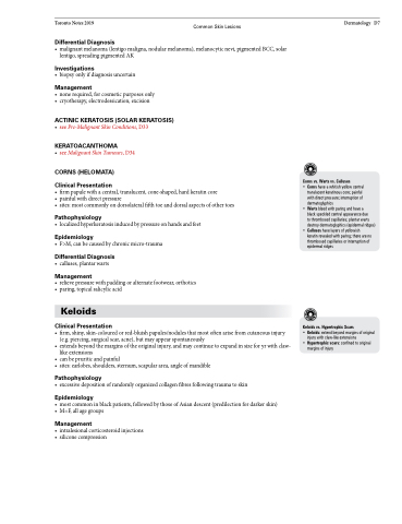
Corns vs. Warts vs. Calluses
• Corns have a whitish yellow central
translucent keratinous core; painful with direct pressure; interruption of dermatoglyphics
• Warts bleed with paring and have a
black speckled central appearance due
to thrombosed capillaries; plantar warts destroy dermatoglyphics (epidermal ridges)
• Calluses have layers of yellowish keratin revealed with paring; there are no thrombosed capillaries or interruption of epidermal ridges
Keloids vs. Hypertrophic Scars
• Keloids: extend beyond margins of original
injury with claw-like extensions
• Hypertrophic scars: confined to original
margins of injury
