
Page 216 - TNFlipTest
P. 216
ER36 Emergency Medicine
Medical Emergencies
Toronto Notes 2019
Electrolyte Disturbances
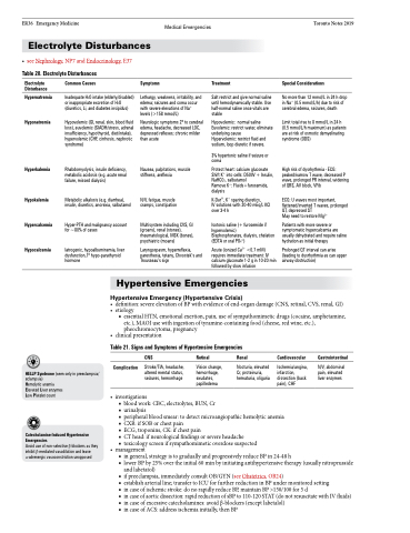
• seeNephrology,NP7andEndocrinology,E37 Table 20. Electrolyte Disturbances
Electrolyte Disturbance
Hypernatremia
Hyponatremia
Hyperkalemia
Hypokalemia
Hypercalcemia
Hypocalcemia
Common Causes
Inadequate H2O intake (elderly/disabled) or inappropriate excretion of H2O (diuretics, Li, and diabetes insipidus)
Hypovolemic (GI, renal, skin, blood fluid loss), euvolemic (SIADH/stress, adrenal insufficiency, hypothyroid, diet/intake), hypervolemic (CHF, cirrhosis, nephrotic syndrome)
Rhabdomyolysis, insulin deficiency, metabolic acidosis (e.g. acute renal failure, missed dialysis)
Metabolic alkalosis (e.g. diarrhea), insulin, diuretics, anorexia, salbutamol
Hyper-PTH and malignancy account for ~80% of cases
Iatrogenic, hypoalbuminemia, liver dysfunction,1o hypo-parathyroid hormone
Symptoms
Lethargy, weakness, irritability, and edema; seizures and coma occur with severe elevations of Na+ levels (>158 mmol/L)
Neurologic symptoms 2o to cerebral edema, headache, decreased LOC, depressed reflexes; chronic milder than acute
Nausea, palpitations, muscle stiffness, areflexia
N/V, fatigue, muscle cramps, constipation
Multisystem including CVS, GI (groans), renal (stones), rheumatological, MSK (bones), psychiatric (moans)
Laryngospasm, hyperreflexia, paresthesia, tetany, Chvostek’s and Trousseau’s sign
Treatment
Salt restrict and give normal saline until hemodynamically stable. Use half-normal saline once vitals are stable
Hypovolemic: normal saline Euvolemic: restrict water, eliminate underlying cause
Hypervolemic: restrict fluid and sodium, loop diuretic if severe.
3% hypertonic saline if seizure or coma
Protect heart: calcium gluconate Shift K+ into cells: D50W + Insulin, NaHC03, salbutamol
Remove K+: Fluids+furosemide, dialysis
K-Dur®, K+ sparing diuretics,
IV solutions with 20-40 mEq/L KCl over 3-4 h
Isotonic saline (+ furosemide if hypervolemic)
Bisphosphonates, dialysis, chelation (EDTA or oral PO43-)
Acute (ionized Ca2+ <0.7 mM) requires immediate treatment: IV calcium gluconate 1-2 g in 10-20 min followed by slow infusion
Special Considerations
No more than 12 mmol/L in 24 h drop in Na+ (0.5 mmol/L/h) due to risk of cerebral edema, seizures, death
Limit total rise to 8 mmol/L in 24 h (0.5 mmol/L/h maximum) as patients are at risk of osmotic demyelinating syndrome (ODS)
High risk of dysrhythmia - ECG: peaked/narrow T wave, decreased P wave, prolonged PR interval, widening of QRS, AV block, VFib
ECG: U waves most important, flattened/inverted T waves, prolonged QT, depressed ST
May need to restore Mg2+
Patients with more severe or symptomatic hypercalcemia are usually dehydrated and require saline hydration as initial therapy
Prolonged QT interval can arise (leading to dysrhythmia as can upper airway obstruction)
Hypertensive Emergencies
Hypertensive Emergency (Hypertensive Crisis)
• definition:severeelevationofBPwithevidenceofend-organdamage(CNS,retinal,CVS,renal,GI) • etiology
■ essential HTN, emotional exertion, pain, use of sympathomimetic drugs (cocaine, amphetamine, etc.), MAOI use with ingestion of tyramine-containing food (cheese, red wine, etc.), pheochromocytoma, pregnancy
• clinicalpresentation
Table 21. Signs and Symptoms of Hypertensive Emergencies
CNS
Stroke/TIA, headache, altered mental status, seizures, hemorrhage
Retinal Renal
Cardiovascular
Ischemia/angina, infarction, dissection (back pain), CHF
Gastrointestinal
N/V, abdominal pain, elevated liver enzymes
HELLP Syndrome (seen only in preeclampsia/ eclampsia)
Hemolytic anemia
Elevated Liver enzymes
Low Platelet count
Catecholamine-Induced Hypertensive Emergencies
Avoid use of non-selective β-blockers as they inhibit β-mediated vasodilation and leave α-adrenergic vasoconstriction unopposed
• investigations
■ blood work: CBC, electrolytes, BUN, Cr
■ urinalysis
■ peripheral blood smear: to detect microangiopathic hemolytic anemia ■ CXR: if SOB or chest pain
■ ECG, troponins, CK: if chest pain
■ CT head: if neurological findings or severe headache
■ toxicology screen if sympathomimetic overdose suspected
Complication
Vision change, hemorrhage, exudates, papilledema
Nocturia, elevated Cr, proteinuria, hematuria, oliguria
• management
■ in general, strategy is to gradually and progressively reduce BP in 24-48 h
■ lower BP by 25% over the initial 60 min by initiating antihypertensive therapy (usually nitroprusside
and labetatol)
■ if preeclampsia, immediately consult OB/GYN (see Obstetrics, OB24)
■ establish arterial line; transfer to ICU for further reduction in BP under monitored setting
■ incaseofischemicstroke:donorapidlyreduceBP,maintainBP>150/100for5d
■ incaseofaorticdissection:rapidreductionofsBPto110-120STAT(donotresuscitatewithIVfluids)
■ in case of excessive catecholamines: avoid β-blockers (except labetalol)
■ in case of ACS: address ischemia initially, then BP
