
Page 353 - TNFlipTest
P. 353
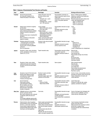
 Toronto Notes 2019 Anatomy Review Table 1. Summary of Gastrointestinal Tract Structure and Function
Gastroenterology G3
Histology and Structural Features
Mucosa: stratified squamous epithelium Submucosa: connective tissue, lymphocytes, plasma cells, nerve cells
Muscularis propria (muscularis externa): inner circular, outer longitudinal muscle
Upper 1/3: striated muscle Middle 1/3: transition zone Lower 1/3: smooth muscle
5 parts Cardia
Fundus Body Antrum Pylorus
4 parts
Superior (5 cm) Descending (7-10 cm) Horizontal (6-8 cm) Ascending (5 cm)
1st part is intraperitoneal; rest is retroperitoneal
Deep red colour
2-4 cm in thickness
Thick and heavy wall
Plicae circulares are large, tall, and closely packed
Has long vasa recta
Scant fat in mesentery
Scant Peyer’s patches
When compared to jejunum Paler pink colour
2-3 cm in thickness
Thin and light walls
Plicae circulares are small and sparse Contains more mesenteric fat
Many Peyer’s patches
Consists of cecum, colon (ascending, transverse, descending, and sigmoid), rectum and anal canal Features include teniae coli, haustra, and omental appendices
Largest internal organ
Composed of 4 lobes (left, right, caudate, quadrate), and divided into 8 segments
Consists of the hepatic ducts (intrahepatic, left, right and common), gallbladder, cystic duct, common bile duct, and ampulla of Vater
4 parts of pancreas: head (includes uncinate process), neck, body, and tail
(Major) pancreatic duct connecting to common bile duct prior to ampulla of Vater
Accessory pancreatic duct connected directly to duodenum
 Organ
Esophagus
Stomach
Duodenum
Jejunum
Ileum
Large Bowel
Liver
Biliary Tract
Pancreas
Function
Muscular tube approximately
40 cm long with a diameter of 2 cm Extends from pharynx to the stomach
Delivers food to intestine for digestion and absorption
Secretes acid, probably to reduce enteric infections/pneumonia; facilitate digestion of protein an absorption of iron/B12
Secretes intrinsic factor to facilitate B12 absorption
Minor contribution to initial protein digestion via pepsin
Modulates enteral pH via secretin
→ decreased gastric acid secretion, increased bicarbonate secretion Secretes CCK to stimulate gallbladder contraction
Site of iron absorption
Absorption of sodium, water, and nutrients (protein, carbohydrates, fat, folic acid, and vitamin A, B, C, D, E, K)
Absorption of sodium, water, nutrients, soluble vitamins (only site of vitamin B12 absorption), and bile salts (entero-hepatic circulation)
Absorption of water (5-10% of total water) Bacteria: further digestion of chyme
and metabolism of undigested CHO
to short chain fatty acids
Formation and storage of feces
Glucose homeostasis
Plasma protein synthesis
Lipid and lipoprotein synthesis Bile acid synthesis and secretion Vitamin A, D, E, K, B12 storage Biotransformation, detoxification Excretion of compounds
Gallbladder functions to store and release bile that is produced in the liver
Bile is used to emulsify fat and is composed of cholesterol, lecithin, bile acids, and bilirubin
CCK stimulates gallbladder emptying while trypsin and chymotrypsin inhibit bile release
Endocrine function: islets of Langerhans produce glucagon, insulin, and somatostatin (from the α, β, and δ cells, respectively) Exocrine function: digestive enzymes are produced including amylase, lipase, trypsin, chymotrypsin, and carboxypeptidase
Blood Supply
Arterial: left gastric artery and left inferior phrenic artery Venous:
Left gastric vein → portal venous system
Esophageal veins → azygos vein → IVC (systemic)
Lesser curvature
Right and left gastric arteries (from celiac trunk)
Greater curvature
Right and left gastro-omental (gastroepiploic) arteries (from gastroduodenal and splenic arteries respectively)
Fundus: short and posterior gastric arteries (from the splenic artery)
Branches of celiac artery and superior mesenteric artery
Superior mesenteric artery
Superior mesenteric artery
Branches of superior and inferior mesenteric arteries
Rectal blood supply: sigmoid, right pudendal, and rectal arteries
2 sources
Portal vein (75-80%) Hepatic artery (20-25%)
Cystic artery
Anterior superior pancreaticoduodenal artery (from the celiac trunk)
Anterior inferior pancreaticoduodenal artery (from the superior mesenteric artery)
Dorsal pancreatic artery (from the splenic artery)
Pancreatic veins drain into the portal, splenic, and superior mesenteric veins
Innervation
Parasympathetic innervation via anterior and posterior gastric nerves (vagal trunks)
Sympathetic innervation via thoracic trunks of the greater splanchnic nerves
Parasympathetic innervation via vagus nerve
Sympathetic innervation via celiac plexus (from T6-T9)
Parasympathetic innervation via vagus nerve
Sympathetic innervation via greater and lesser splanchnic nerves
Parasympathetic innervation via fibres of the posterior vagal trunk Sympathetic innervation via fibres of T8-T10
Same as jejunum
Parasympathetic innervation via vagus nerve
Sympathetic innervation via greater and lesser splanchnic nerves
Parasympathetic innervation via fibres of the anterior and posterior vagal trunks
Sympathetic innervation via fibres of the celiac plexus
Parasympathetic innervation via vagus nerve
Sympathetic and visceral innervation via celiac nerve plexus
Somatic afferent fibres via right phrenic nerve
Parasympathetic innervation via vagus nerve
Sympathetic innervation via abdominopelvic splanchnic nerves
