
Page 408 - TNFlipTest
P. 408
GS6 General Surgery and Thoracic Surgery Differential Diagnoses of Common Presentations Toronto Notes 2019
Indications for Urgent Operation
IHOP Ischemia Hemorrhage Obstruction Perforation
Overt bleeding: obvious hematemesis, hematochezia or melena per rectum (i.e. visible to naked eye)
Occult bleeding: bleeding per rectum is not obvious to naked eye (e.g. positive guaiac FOBT)
Obscure bleeding: bleeding with no identifiable source after colonoscopy and endoscopy (source usually in small bowel). Can be either overt or occult
Transfusion Strategies for Acute Upper Gastrointestinal Bleeding
NEJM 2013;368:11-21
Recent study by Villanueva et al., demonstrates that a restrictive transfusion strategy (transfusion with hemoglobin below 70 g/L) significantly improves outcomes in patients with acute UGIB, compared
to a liberal transfusion strategy (transfusion with hemoglobin below 90 g/L). Refer to study for details.
Gastrointestinal Bleeding
• seeGastroenterology,G25
Indications for Surgery
• failureofmedicalmanagement
• exsanguinatinghemorrhage:hemodynamicinstabilitydespitevigorousresuscitation • recurrenthemorrhagewithuptotwoattemptsofendoscopichemostasis
• prolongedbleedingwithtransfusionrequirement>3units
• bleeding at rate >1 unit/8 h
Surgical Management of GI Bleeding
• UGIB
■ bleeding from a source proximal to the ligament of Treitz
■ oftenpresentswithhematemesisandmelenaunlessverybrisk(thencanpresentwithhematochezia) ■ initial management with endoscopy; if fails, then consider surgical management appropriate to
etiology
■ PUD accounts for approximately 55% of severe UGIB
• LGIB
■ bleeding from a source distal to the ligament of Treitz
■ often presents with BRBPR unless proximal to transverse colon
◆ may occasionally present with melena
■ initial management with colonoscopy to detect and potentially stop source of bleeding
■ 75% of patients will spontaneously stop bleeding, however if bleeding continues barium enema
should NOT be performed
■ angiography or RBC scan to determine source as indicated
■ surgery indicated if bleeding is persistent - aimed at resection of area containing source of bleeding ■ obscure bleed may require blind total colectomy if the source is not found
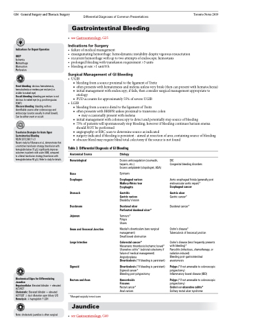
Table 3. Differential Diagnosis of GI Bleeding
Biochemical Signs for Differentiating Jaundice
Hepatocellular: Elevated bilirubin + elevated ALT/AST
Cholestatic: Elevated bilirubin + elevated ALP/GGT ± duct dilatation upon biliary U/S Hemolysis: haptoglobin LDH
Anatomical Source
Hematological
Nose Esophagus
Stomach
Duodenum Jejunum
Ileum and Ileocecal Junction Large Intestine
Sigmoid
Rectum and Anus
*Managed surgically in most cases
Jaundice
Etiology
Excess anticoagulation (coumadin, heparin, etc.)
Excess antiplatelet (clopidogrel, ASA)
Epistaxis
Esophageal varices Mallory-Weiss tear Esophagitis
Gastritis
Gastric varices Dieulafoy’s lesion
Duodenal ulcer
Perforated duodenal ulcer*
Tumours* Polyps Ulcers
Meckel’s diverticulum (rare surgical management)
Small bowel obstruction
Colorectal cancer*
Mesenteric thrombosis/ischemic bowel* Ulcerative colitis* (subtotal colectomy if failure of medical management) Angiodysplasia
Diverticulosis (*if bleeding is persistent)
Diverticulosis (*if bleeding is persistent) Sigmoid cancer*
Bleeding post-polypectomy
Hemorrhoids Fissures Rectal cancer* Anal varices
DIC
Congenital bleeding disorders
Aorto-esophageal fistula (generally post endovascular aortic repair)* Esophageal cancer
Gastric ulcer
Gastric cancer* Duodenal cancer*
Crohn’s disease*
Tuberculosis of ileocecal junction
Crohn’s disease (less frequently presents with bleeding)*
Pancolitis (infectious, chemotherapy, or radiation induced)
Bleeding post-gastrointestinal anastomosis
Polyps (*if not amenable to colonoscopic polypectomy)
Inflammatory bowel disease (IBD)
Polyps (*if not amenable to colonoscopic polypectomy)
Crohn’s or ulcerative colitis*
Solitary rectal ulcer syndrome
Note: cholestatic jaundice is often surgical
• seeGastroenterology,G40
