
Page 406 - TNFlipTest
P. 406
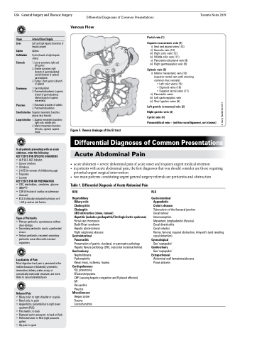
 GS4 General Surgery and Thoracic Surgery Differential Diagnoses of Common Presentations Toronto Notes 2019 Venous Flow
    Organ Liver
Spleen Gallbladder
Stomach
Duodenum
Pancreas Small intestine Largeintestine
Arterial Blood Supply
Left and right hepatic (branches of hepatic proper)
Splenic 426 Cystic (branch of right hepatic 1
artery) 3
1. Lesser curvature: right and 5
Portal vein (1)
Superior mesenteric vein (7)
i) Ileal and jejunal veins (13) ii) Ileocolic vein (14)
iii) Right colic vein (12) iv) Middle colic vein (11)
v) Pancreaticoduodenal vein (8) vi) Right gastroepiploic vein (9)
Splenic vein (5)
i) Inferior mesenteric vein (10) (superior rectal vein until crossing common iliac vessels)
      left gastric
2. Greater curvature: right
7 10
8 9
11
2. Pancreaticoduodenals (superior 12 15
branch of gastroduodenal,
inferior branch of superior 13 mesenteric) 16
1. Pancreatic branches of splenic 14
2. Pancreaticoduodenals 17
  (branch of gastroduodenal) and left (branch of splenic) gastroepiploic
   3. Fundus: short gastrics (branch of splenic)
• Left colic veins (15)
• Sigmoid veins (16)
• Superior rectal veins (17)
Pancreatic veins
Left gastroepiploic vein Short gastric veins (6)
  1. Gastroduodenal
     ii) iii) iv)
        Superior mesenteric branches: jejunal, ileal, ileocolic
1.Superiormesentericbranches: right colic, middle colic
2. Inferior mesenteric branches: left colic, sigmoid, superior rectal
Left gastric (coronary) vein (2)
Right gastric vein (3)
Cystic vein (4)
Paraumbilical vein – (within round ligament, not shown)
Figure 5. Venous drainage of the GI tract
   In all patients presenting with an acute abdomen, order the following:
KEY TESTS FOR SPECIFIC DIAGNOSIS • ALP, ALT, AST, bilirubin
• Lipase/ amylase
• Urinalysis
• β-hCG(inwomenofchildbearingage) • Troponins
• Lactate
KEY TESTS FOR OR PREPARATION
• CBC, electrolytes, creatinine, glucose
• INR/PTT
• CXR (if history of cardiac or pulmonary
disease)
• ECG if clinically indicated by history or if
>69 yr and no risk factors
Types of Peritonitis
• Primary peritonitis: spontaneous without clear etiology
• Secondary peritonitis: due to a perforated viscus
• Tertiary peritonitis: recurrent secondary peritonitis more often with resistant organisms
Localization of Pain
Most digestive tract pain is perceived in the midline because of bilaterally symmetric innervation; kidney, ureter, ovary, or somatically innervated structures are more likely to cause lateralized pain
Referred Pain
• Biliary colic: to right shoulder or scapula
• Renal colic: to groin
• Appendicitis: periumbilical to right lower
quadrant (RLQ)
• Pancreatitis: to back
• Ruptured aortic aneurysm: to back or flank
• Perforated ulcer: to RLQ (right paracolic
gutter)
• Hip pain: to groin
Differential Diagnoses of Common Presentations
Acute Abdominal Pain
• acuteabdomen=severeabdominalpainofacuteonsetandrequiresurgentmedicalattention
• inpatientswithacuteabdominalpain,thefirstdiagnosesthatyoushouldconsiderarethoserequiring
potential urgent surgical intervention
• twomainpatternsconstitutingurgentgeneralsurgeryreferralsareperitonitisandobstruction
 Table 1. Differential Diagnosis of Acute Abdominal Pain
RUQ
Hepatobiliary
Biliary colic
Cholecystitis
Cholangitis
CBD obstruction (stone, tumour)
Hepatitis (includes perihepatitis/Fitz-Hugh-Curtis syndrome) Portal vein thrombosis
Budd-Chiari syndrome Hepatic abscess/mass Right subphrenic abscess
Gastrointestinal
Pancreatitis
Presentation of gastric, duodenal, or pancreatic pathology Hepatic flexure pathology (CRC, subcostal incisional hernia)
Genitourinary
Nephrolithiasis
Pyelonephritis
Renal: mass, ischemia, trauma
Cardiopulmonary
RLL pneumonia
Effusion/empyema
CHF (causing hepatic congestion and R pleural effusion) MI
Pericarditis
Pleuritis
Miscellaneous
Herpes zoster Trauma Costochondritis
RLQ
Gastrointestinal
Appendicitis
Crohn’s disease
Tuberculosis of the ileocecal junction Cecal tumour
Intussusception
Mesenteric lymphadenitis (Yersinia)
Cecal diverticulitis
Cecal volvulus
Hernia: femoral, inguinal obstruction, Amyand’s (and resulting cecal distention)
Gynecological
See ‘suprapubic’
Genitourinary
See ‘suprapubic’
Extraperitoneal
Abdominal wall hematoma/abscess Psoas abscess
        © Carly Vanderlee 2011
