
Page 445 - TNFlipTest
P. 445
Toronto Notes 2019
Liver
General Surgery and Thoracic Surgery GS43
Liver Cysts
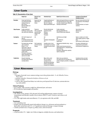
Table 18. Characteristics of Liver Cysts
Description
Clinical Features
Investigations
Treatment
Complications
Simple Cysts
Contain clear fluid that do not communicate with the intrahepatic biliary tree Most common
May have multiple cysts Always benign
Usually asymptomatic
Dull RUQ pain, bloating, and/or early satiety when symptomatic
U/S: Used for diagnosis and follow-up
CT: well demarcated lesion that does not enhance with contrast
Not required unless very large and/or symptomatic
Monitor if >4 cm Laparoscopic or open cyst wall removal (unroofing) is established treatment and Is usually curative Percutaneous drainage and ethanol sclerotherapy also an option, but not curative
Intracystic hemorrhage
Cyst rupture which can lead to secondary infection
Polycystic Liver Disease
Several cysts that replace much of the liver
Progressive
50% associated with polycystic kidney disease (if over
age 60)
U/S
Only if symptomatic partial liver resection drainage
Choledochal Cysts
Congenital malformations of pancreaticobiliary tree
High risk of malignancy Majority present before age 10
Recurrent abdominal pain Intermittent jaundice RUQ mass
Cholangitis
Pancreatitis
U/S
CT
Transhepatic cholangiography LFTs
Complete excision of cysts liver transplant if cyst involves intrahepatic bile ducts (Caroli’s disease)
Biliary cirrhosis, portal
HTN, cyst rupture, or cholangiocarcinoma Abnormal pancreaticobiliary junction is associated with increased risk of malignancy
Hydatid (Cystic Echinococcosis)
Infection with parasite Echinococcus granulosus Associated with exposure to dogs, sheep,
and cattle in Southern Europe, Middle East, Australasia, South America
Usually asymptomatic
May have palpable RUQ mass Chronic RUQ pain when symptomatic
Anti-Echinococcus Ab (IgG) U/S
CT: calcified mass
Needle biopsy
Albendazole (anti-helminthic drug) – cure up to 30%
Surgical: radical (total pericystectomy or partial hepatectomy) vs. conservative (open endocystectomy)
Percutaneous: PAIR (puncture, aspiration, injection, reaspiration)
Inferior vena cava compression
Cyst rupture which can cause biliary colic, jaundice, cholangitis, pancreatitis, or anaphylactic reaction
Cystadenoma (Premalignant)/ Cystadenocarcinoma
Rare cystic tumours that occur in the liver parenchyma or the extrahepatic bile ducts
Cystadenocarcinoma is an invasive carcinoma
Upper abdominal mass Abdominal pain Anorexia
Appear as complex cysts: internal septae, papillary projections, irregular lining
Need histology for definite diagnosis
All complex, multiloculated cysts (except echinococcal) should be excised because of malignancy risk
Cystadenocarcinoma can invade adjacent tissues and metastasize
Liver Abscesses
Etiology
• types
■ pyogenic (bacterial): most common etiology; most often polymicrobial – E. coli, Klebsiella, Proteus,
Strep. milleri
■ parasitic (amoebic): Entamoeba histolytica, Echinococcal cyst
■ fungal: Candida
■ sources: direct spread from biliary tract infection, portal spread from GI infection, systemicinfection
(e.g. endocarditis)
Clinical Features
• fever,malaise,chills,anorexia,weightloss,abdominalpain,andnausea • RUQtenderness,hepatomegaly,andjaundice
Investigations
• CBC(leukocytosis,anemia),LFTs(elevatedALPandhypoalbuminemiacommon;elevated transaminases and bilirubin variable), blood cultures, INR/PTT, and E. histolytica and echinoccocal serology
• U/S,CXR(rightbasilaratelectasis/effusion),CT,cystaspirationwithC&S,andMRI
Treatment
• treatunderlyingcause
• pyogenicabscessesgenerallytreatedwithantibiotictherapy(e.g.ceftriaxoneandmetronidazoleor
piperacillin-tazobactam) and U/S- or CT-guided percutaneous drainage or surgical drainage
• considerpotentialsourceofsepsis(e.g.biliarysource,infectedtumour)
Prognosis
• overallmortality15%–higherrateifdelayindiagnosis,multipleabscesses,andmalnutrition
