Page 459 - TNFlipTest
P. 459
 Toronto Notes 2019 Breast General Surgery and Thoracic Surgery GS57
Diagnostic Procedures
• "tripletest"fordiagnosisofbreastcancer:clinicalbreastexam,imaging(U/Sfor<30yr,mammography + U/S for >30 yr), pathology (biopsy)
• needleaspiration:forpalpablecysticlesions;sendfluidforcytologyifbloodorcystdoesnotcompletely resolve
• U/Sormammographyguidedcoreneedlebiopsy(mostcommon)
• excisionalbiopsy:onlyperformedassecondchoicetocoreneedlebiopsy;shouldnotbedonefor
diagnosis if possible
Genetic Screening
• considertestingforBRCA1/2if:
■ patient diagnosed with breast AND ovarian cancer ■ strong family history of breast/ovarian cancer
■ family history of male breast cancer
■ young patient (<35 yr)
■ bilateral breast cancer in patients <50 yr
Staging
• patientsareassignedaclinicalstagepre-operatively(cTNM);followingsurgerythepathologicstageis determined (pTNM)
• clinical
■ tumour size by palpation, mammogram, U/S and/or MRI
■ nodal involvement by palpation, imaging
■ metastasis by physical exam, CXR, and abdominal U/S (or CT chest/abdomen/pelvis), bone scan
(usually done post-operative if node-positive disease)
• pathological
■ tumour size and type
■ grade: modified Bloom and Richardson score (I to III) – histologic, nuclear, and mitotic grade
■ number of axillary nodes positive for malignancy out of total nodes resected, extranodal extension,
and sentinel lymph node biopsy (SLNB) positive/negative
■ tumour biology: estrogen receptor (ER), progesterone receptor (PR), and HER2/neu oncogene
status
■ margins: for invasive breast cancer negative margin is sufficient, for DCIS prefer 2 mm margin ■ lymphovascular invasion (LVI)
■ extensive in situ component (EIC): DCIS in surrounding tissue
■ involvement of dermal lymphatics (inflammatory) – automatically Stage IIIb
Diagnostic mammography is indicated in all patients, even in women <50 yr
Phyllodes tumours are rare fibroepithelial breast tumours that can be benign or malignant that mostly affect women from 35-55 yr
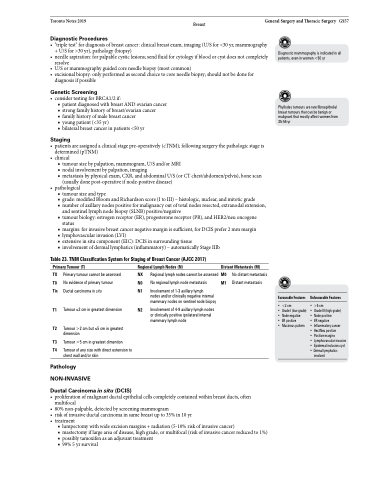
    Table 23. TNM Classification System for Staging of Breast Cancer (AJCC 2017)
Primary Tumour (T) Regional Lymph Nodes (N)
Distant Metastasis (M)
  TX Primary tumour cannot be assessed NX T0 No evidence of primary tumour N0 Tis Ductal carcinoma in situ N1
T1 Tumour ≤2 cm in greatest dimension N2
T2 Tumour >2 cm but ≤5 cm in greatest
dimension
T3 Tumour >5 cm in greatest dimention
T4 Tumour of any size with direct extension to chest wall and/or skin
Pathology NON-INVASIVE
Regional lymph nodes cannot be assessed M0 No regional lymph node metastasis M1
Involvement of 1-3 axillary lymph
nodes and/or clinically negative internal mammary nodes on sentinel node biopsy
Involvement of 4-9 axillary lymph nodes or clinically positive ipsilateral internal mammary lymph node
No distant metastasis Distant metastasis
  Favourable Features
• <2 cm
• Grade I (low grade)
• Node negative
• ER positive
• Mucinous pattern
Unfavourable Features
• >5 cm
• Grade III (high grade)
• Node positive
• ER negative
• Inflammatory cancer
• Her2Neupositive
• Positivemargins
• Lymphovascularinvasion • Epidermalinclusioncyst • Dermal lymphatics
involved
 Ductal Carcinoma in situ (DCIS)
• proliferationofmalignantductalepithelialcellscompletelycontainedwithinbreastducts,often
multifocal
• 80%non-palpable,detectedbyscreeningmammogram
• riskofinvasiveductalcarcinomainsamebreastupto35%in10yr
• treatment
■ lumpectomy with wide excision margins + radiation (5-10% risk of invasive cancer)
■ mastectomy if large area of disease, high grade, or multifocal (risk of invasive cancer reduced to 1%) ■ possibly tamoxifen as an adjuvant treatment
■ 99% 5 yr survival


