
Page 457 - TNFlipTest
P. 457
Toronto Notes 2019
Breast
General Surgery and Thoracic Surgery GS55
Breast
Left bronchomediastinal trunk
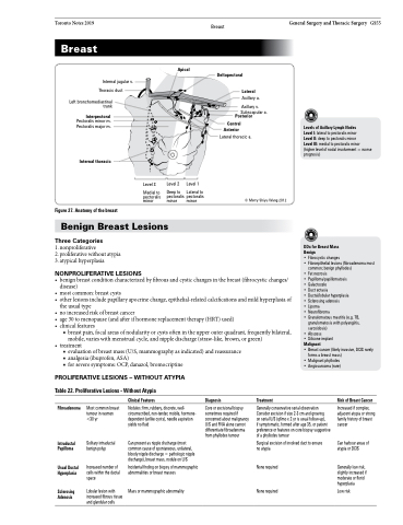
Apical
Internal jugular v. Thoracic duct
Deltopectoral Lateral
Axillary a.
Axillary v.
Subscapular a.
Interpectoral
Posterior Central
Pectoralis minor m. Pectoralis major m.
Anterior
Lateral thoracic a.
© Merry Shiyu Wang 2012
Levels of Axillary Lymph Nodes
Level I: lateral to pectoralis minor
Level II: deep to pectoralis minor
Level III: medial to pectoralis minor (higher level of nodal involvement = worse prognosis)
Internal thoracic
Figure 27. Anatomy of the breast
Level 3
Medial to pectoralis minor
Level 2
Level 1
Lateral to pectoralis pectoralis
Deep to minor
minor
Benign Breast Lesions
Three Categories
1. nonproliferative
2. proliferative without atypia 3. atypical hyperplasia
NONPROLIFERATIVE LESIONS
DDx for Breast Mass
Benign
• Fibrocystic changes
• Fibroepithelial lesions (fibroadenoma most
common; benign phyllodes)
• Fat necrosis
• Papilloma/papillomatosis
• Galactocele
• Duct ectasia
• Ductal/lobular hyperplasia
• Sclerosing adenosis
• Lipoma
• Neurofibroma
• Granulomatous mastitis (e.g. TB,
granulomatosis with polyangiitis,
sarcoidosis)
• Abscess
• Silicone implant Malignant
• Breast cancer (likely invasive, DCIS rarely
forms a breast mass)
• Malignant phyllodes
• Angiosarcoma (rare)
• benignbreastconditioncharacterizedbyfibrousandcysticchangesinthebreast(fibrocysticchanges/ disease)
• mostcommon:breastcysts
• otherlesionsincludepapillaryapocrinechange,epithelial-relatedcalcificationsandmildhyperplasiaof
the usual type
• noincreasedriskofbreastcancer
• age30tomenopause(andafterifhormonereplacementtherapy(HRT)used)
• clinicalfeatures
■ breast pain, focal areas of nodularity or cysts often in the upper outer quadrant, frequently bilateral, mobile, varies with menstrual cycle, and nipple discharge (straw-like, brown, or green)
• treatment
■ evaluation of breast mass (U/S, mammography as indicated) and reassurance ■ analgesia (ibuprofen, ASA)
■ for severe symptoms: OCP, danazol, bromocriptine
PROLIFERATIVE LESIONS – WITHOUT ATYPIA
Table 22. Proliferative Lesions - Without Atypia
Fibroadenoma
Intraductal Papilloma
Usual Ductal Hyperplasia
Sclerosing Adenosis
Most common breast tumour in women <30 yr
Solitary intraductal benign polyp
Increased number of cells within the ductal space
Lobular lesion with increased fibrous tissue and glandular cells
Clinical Features
Nodules: firm, rubbery, discrete, well- circumscribed, non-tender, mobile, hormone- dependent (unlike cysts), needle aspiration yields no fluid
Can present as nipple discharge (most common cause of spontaneous, unilateral, bloody nipple discharge = pathologic nipple discharge), breast mass, nodule on U/S
Incidental finding on biopsy of mammographic abnormalities or breast masses
Mass or mammographic abnormality
Diagnosis
Core or excisional biopsy sometimes required if concerned about malignancy U/S and FNA alone cannot differentiate fibroadenoma from phyllodes tumour
Treatment
Generally conservative serial observation Consider excision if size 2-3 cm and growing
on serial U/S (q6mo x 2 yr is usual follow-up),
if symptomatic, formed after age 35, or patient preference or features on core biopsy suggestive of a phyllodes tumour
Surgical excision of involved duct to ensure no atypia
None required
Risk of Breast Cancer
Increased if complex, adjacent atypia or strong family history of breast cancer
Can harbour areas of atypia or DCIS
Generally low risk, slightly increased if moderate or florid hyperplasia
Low risk
None required
