
Page 544 - TNFlipTest
P. 544
H4 Hematology
Basics of Hematology
Toronto Notes 2019
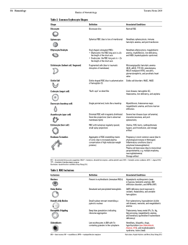
Table 2. Common Erythrocyte Shapes
Shape
Discocyte Spherocyte Elliptocyte/Ovalcyte
Schistocyte (helmet cell, fragment)
Sickle Cell
Codocyte (target cell)
Dacrocyte (teardrop cell) Acanthocyte (spur cell) Echinocyte (burr cell)
Rouleaux Formation
Definition
Biconcave disc
Spherical RBC (due to loss of membrane)
Oval-shaped, elongated RBCs
• Elliptocytes: the RBC long axis is ≥2x
the length of the short axis
• Ovalocytes: the RBC long axis is <2x
the length of the short axis
Fragmented cells (due to traumatic disruption of membrane)
Sickle-shaped RBC (due to polymerization of hemoglobin S)
“Bull’s eye” on dried film
Single pointed end, looks like a teardrop
Distorted RBC with irregularly distributed thorn-like projections (due to abnormal membrane lipids)
RBC with numerous regularly spaced, small spiny projections
Aggregates of RBC resembling stacks of coins (due to increased plasma concentration of high molecular weight proteins)
Associated Conditions
Normal RBC
Hereditary spherocytosis, immune hemolytic anemia, and post-transfusion
Hereditary elliptocytosis, megaloblastic anemia, myelofibrosis, iron-deficiency, and MDS (myelodysplastic syndrome)
Microangiopathic hemolytic anemia (HUS, aHUS, TTP, DIC, preeclampsia, HELLP, malignant HTN), vasculitis, glomerulonephritis, and prosthetic heart valve
Sickle cell disorders: HbSC, HbSS
Liver disease, hemoglobin SC, thalassemia, iron deficiency, and asplenia
Myelofibrosis, thalassemia major, megaloblastic anemia, and bone marrow infiltration
Severe liver disease (spur cell anemia), starvation/anorexia, and post- splenectomy
Uremia, HUS, burns, cardiopulmonary bypass, post-transfusion, and storage artifact
Pregnancy is most common cause (due to physiological increase in fibrinogen) Inflammatory conditions (due to polyclonal immunoglobulins)
Plasma cell dyscrasias (due to monoclonal paraproteinemia, e.g. multiple myeloma, macroglobulinemia)
Storage artifact
DIC = disseminated intravascular coagulation; HELLP = hemolysis, elevated liver enzymes, and low platelet count; HUS = hemolytic uremic syndrome; aHUS = atypical HUS; TTP = thrombotic thrombocytopenic purpura
Illustrations: Ayalah Hutchins and Merry Shiyu Wang 2012
Table 3. RBC Inclusions
Inclusions
Nucleus Heinz Bodies
Howell-Jolly Bodies Basophilic Stippling
Sideroblasts
Definition
Present in erythroblasts (immature RBCs) Denatured and precipitated hemoglobin
Small nuclear remnant resembling a pyknotic nucleus
Deep blue granulations indicating ribosome aggregation
Late erythrocytes in BM with Fe containing granules in the cytoplasm
Associated Conditions
Hyperplastic erythropoiesis (seen
in hypoxia, hemolytic anemia), BM infiltration disorders, and MPNs (MF)
G6PD deficiency (post-exposure to oxidant), thalassemia, and unstable hemoglobins
Post-splenectomy, hyposplenism (sickle cell disease), neonates, and megaloblastic anemia
Thalassemia, heavy metal (Pb, Zn, Ag, Hg) poisoning, megaloblastic anemia, and hereditary (pyrimidine 5’nucleotidase deficiency)
Hereditary, idiopathic, drugs, hypothyroidism (see Sideroblastic Anemia, H16), and myelodysplastic syndrome, toxins (lead)
BM = bone marrow; MF = myelofibrosis; MPN = myeloproliferative neoplasm
Illustrations: Ayalah Hutchins and Merry Shiyu Wang 2012
