
Page 698 - TNFlipTest
P. 698
MI30 Medical Imaging
Breast Imaging Toronto Notes 2019 Breast Imaging
Modalities
Mammography
Description
• x-rayimagingofthebreastsforscreeninginasymptomaticpatients,ordiagnosisofclinically-detected or screening-detected abnormalities (see General Surgery, GS55)
• routineevaluationinvolvestwostandardviews:cranio-caudalandmedial-lateral-oblique
Indications
• screening
■ beginscreeningfromage50q2-3yr
■ no strong data to support screening >70 yr, but may continue screening if in good general health ■ if <50, screening is only recommended for those with high risk of breast cancer
■ screening detects 2-8 cancers/1,000 women screened
• surveillance
■ follow-up of women with previous breast cancer
• diagnostic:includesmammographywithspecialviewsand/orultrasound
■ workup of an abnormality that may be suggestive of breast cancer including a lump or thickening,
localized nodularity, dimpling or contour deformity, a persistent focal area of pain, overlying skin
changes, and spontaneous serous or sanguinous nipple discharge from a single duct
■ women with abnormal screening mammograms
■ suspected complications of breast implants
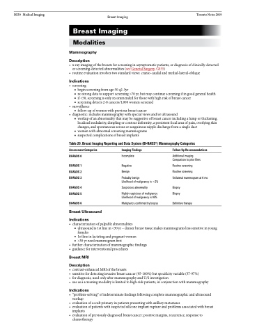
Table 20. Breast Imaging Reporting and Data System (BI-RADS®) Mammography Categories
Assessment Categories
BI-RADS 0
BI-RADS 1 BI-RADS 2 BI-RADS 3
BI-RADS 4 BI-RADS 5
BI-RADS 6
Breast Ultrasound
Indications
Imaging Findings
Incomplete
Negative Benign
Probably benign
Likelihood of malignancy is <2%
Suspicious abnormality
Highly suspicious of malignancy Likelihood of malignancy is 95%
Malignancy confirmed by biopsy
Follow-Up Recommendations
Additional imaging Comparison to prior films
Routine screening
Routine screening
Unilateral mammogram at 6 mo
Biopsy Biopsy
Definitive therapy
• characterizationofpalpableabnormalities
■ ultrasound is 1st line in <30 yr – denser breast tissue makes mammograms less sensitive in young
females
■ 1st line in lactating and pregnant women ■ >30yrneedmammogramfirst
• furthercharacterizationofmammographicfindings • guidanceforinterventionalprocedures
Breast MRI
Description
• contrast-enhancedMRIofthebreasts
• sensitivefordetectinginvasivebreastcancer(95-100%)butspecificityvariable(37-97%)
• fordiagnosis,usedonlyaftermammographyandU/Sinvestigation
• useasascreeningmodalityislimitedtohigh-riskpatients,inconjunctionwithmammography
Indications
• “problem-solving”ofindeterminatefindingsfollowingcompletemammographicandultrasound workup
• evaluationofoccultprimaryinpatientspresentingwithaxillarymetastases
• evaluationofpatientswithsuspectedsiliconeimplantruptureandproblemsassociatedwithbreast
implants
• evaluationofpreviouslydiagnosedbreastcancer:positivemargins,recurrence,responseto
chemotherapy
