
Page 699 - TNFlipTest
P. 699
Toronto Notes 2019 Breast Imaging Medical Imaging MI31
• high-riskscreening
■ known BRCA1 or BRCA2 mutation, or other gene predisposing to breast cancer, or untested first-
degree relative of a carrier of such a gene mutation
■ family history consistent with a hereditary breast cancer syndrome and/or estimated personal
lifetime cancer risk >25%
■ high-risk marker on prior biopsy (atypical ductal hyperplasia, atypical lobular hyperplasia, lobular
carcinoma in situ)
■ radiation therapy to chest (before age 30)
Breast Interventional Procedures
Description
• includesfineneedleaspiratebiopsy,coreneedlebiopsy,stereotacticbiopsy,MRIguidedbiopsy,abscess drainage, and cyst aspiration (see General Surgery, GS59)
Indications
• cysticmass:complexcyst,symptomatic,suspectedabscess
• solidmass:confirmdiagnosisofalesionsuspiciousformalignancy(BI-RADS®Category4or5)
• suspiciouscalcifications:confirmdiagnosisofalesionsuspiciousformalignancy(BI-RADS®Category4
or 5) – stereotactic biopsy
• initialpercutaneousbiopsyprocedurethatwasinsufficientordiscordantwithimaging • presurgicalwirelocalizationofalesion
Breast Findings
Breast Masses
• definition:aspace-occupyinglesionseenintwodifferentprojections;ifseeninonlyasingleprojection it should be called an “asymmetry” until its three-dimensionality is confirmed
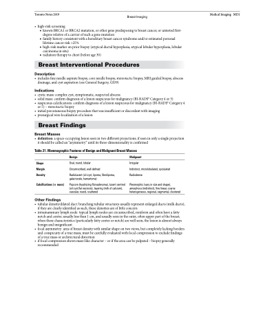
Table 21. Mammographic Features of Benign and Malignant Breast Masses
Shape Margin Density
Calcifications (± mass)
Other Findings
Benign
Oval, round, lobular Circumscribed, well-defined
Radiolucent (oil cyst, lipoma, fibrolipoma, galactocele, hamartoma)
Popcorn (hyalinizing fibroadenoma), lucent centred (oil cyst/fat necrosis), layering (milk of calcium), vascular, round, scattered
Malignant
Irregular
Indistinct, microlobulated, spiculated Radiodense
Pleomorphic (vary in size and shape), amorphous (indistinct), fine linear, coarse heterogeneous, regional, segmental, clustered
• tubulardensity/dilatedduct:branchingtubularstructuresusuallyrepresentenlargedducts(milkducts); if they are clearly identified as such, these densities are of little concern
• intramammarylymphnode:typicallymphnodesarecircumscribed,reniformandoftenhaveafatty notch and centre; usually less than 1 cm, and usually seen in the outer, often upper part of the breast; when these characteristics (particularly fatty centre or notch) are well seen, the lesion is almost always benign and insignificant
• focalasymmetry:areaofbreastdensitywithsimilarshapeontwoviews,butcompletelylackingborders and conspicuity of a true mass; must be carefully evaluated with focal compression to exclude findings of a true mass or architectural distortion
• iffocalcompressionshowsmass-likecharacter–oriftheareacanbepalpated–biopsygenerally recommended
