Page 706 - TNFlipTest
P. 706
 NP6 Nephrology
Assessment of Renal Function
Toronto Notes 2019
   Estimating Urine Osmolality
Last 2 digits of the specific gravity x 30 = urine osmolality approximately
(e.g. specific gravity of 1.020 = 600 mOsm)
Urinalysis
• usedipstickinfreshlyvoidedurinespecimentoassessthefollowing:
1 . Specific Gravity
• ratioofthemassofequalvolumesofurine/H2O
• rangeis1.001-1.030
• values<1.010reflectdiluteurine,values>1.020reflectconcentratedurine • valueusually1.010(isosthenuria:samespecificgravityasplasma)inESRD
2 . pH
• urinepHisnormallybetween4.5-7.0;ifpersistentlyalkaline,consider ■ RTA
■ UTI with urease-producing bacteria (e.g. Proteus)
3 . Glucose
• freelyfilteredatglomerulusandreabsorbedinproximaltubule • causesofglucosuriainclude:
1. hyperglycemia >9-11.1 mmol/L leads to filtration that exceeds tubular resorption capacity
2. increased GFR (e.g. pregnancy - the proximal convoluted tubule is unable to reabsorb the glucose
and amino acids)
3. proximal tubule dysfunction (e.g. Fanconi’s syndrome)
4. sodium-glucose cotransporter 2 (SGLT2) inhibitors (also known as -flozin drugs) which are prescribed
for DM2, lower the threshold for glucosuria by preventing glucose reabsorption from the filtrate
4 . Protein
• dipstickonlydetectsalbumin;otherproteins(e.g.Bence-Jones,Ig,Tamm-Horsfall)maybemissed
• microalbuminuria(morningACRof2.0-20mg/mmol)isnotdetectedbystandarddipstick,greater
than these ranges would be macroalbuminuria (see Diabetes, NP11) • gold standard: 24 h timed urine collection for total protein
5 . Leukocyte Esterase
• enzymefoundinWBCanddetectedbydipstick
• presenceofWBCsindicatesinfection(e.g.UTI)orinflammation(e.g.AIN)
6 . Nitrites
• endogenousnitratesinurineareconvertedtonitritesbysomebacteria(mostcommonlyE.coli) • highspecificitybutlowsensitivityforUTI
7 . Ketones
• positiveinalcoholic/diabeticketoacidosis,prolongedstarvation,fasting
8 . Hemoglobin
• positiveinhemoglobinuria(hemolysis),myoglobinuria(rhabdomyolysis),andtruehematuria(RBCs seen on microscopy)
  24 h Urine Collection
• Discard first morning specimen
• Collect all subsequent urine for the
next 24 h
• Refrigerate between voids
• Collect second morning specimen and take
to lab immediately
Positive dipstick for leukocyte esterase and nitrites is highly specific for diagnosing a UTI
Nitrite Negative Bacteria
• Enterococci • Staphylococci
Nitrite Positive Bacteria
• Enterobacteriacae (e.g. E. coli)
       Urine Microscopy
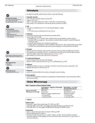
Table 2. Comparison of Urinary Sediment Findings
  Any one or more of the following seen on microscopy
1 . CELLS
Erythrocytes
Active Sediment = Suggestive of Parenchymal Kidney Disease
Red cell casts
White cell casts
Muddy-brown granular or epithelial cell casts >2 red cells per HPF
>4 white cells per HPF
Bland Sediment = Less Likely Parenchymal Kidney Disease
Only hyaline casts
Small quantities of crystals Small amount of bacteria <2 red cells per HPF
<4 white cells per HPF
 • hematuria=greaterthannormalrangeof2-3RBCsperHPF
• dysmorphicRBCsand/orRBCcastssuggestglomerularbleeding(e.g.proliferativeGN) • isomorphicRBCs,nocastssuggestextraglomerularbleeding(e.g.bladdercancer)
Leukocytes
• pyuria=greaterthanupperlimitofnormal:5WBCsperHPF
• indicatesinflammationorinfection
• ifpersistentsterilepyuriapresent(i.e.negativeculture),consider:chronicurethritis,prostatitis,
interstitial nephritis, calculi, papillary necrosis, renal tuberculosis, viral infections, N. gonorrhoeae, C. trachomatis infection


