
Page 713 - TNFlipTest
P. 713
Toronto Notes 2019 Electrolyte Disorders
Hyperkalemia
• serum[K+]>5.0mEq/L
Signs and Symptoms
• usuallyasymptomaticbutmaydevelopnausea,palpitations,muscleweakness,musclestiffness, paresthesias, areflexia, ascending paralysis, and hypoventilation
Nephrology NP13
• impairedrenalammoniagenesisandmetabolicacidosis
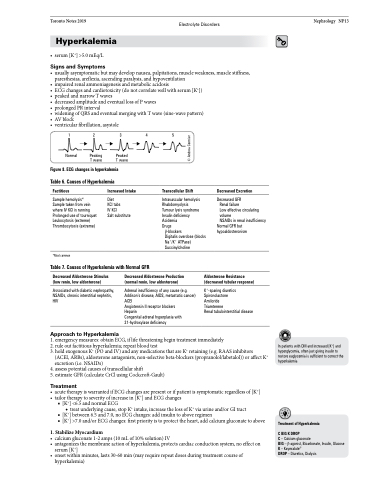
• ECGchangesandcardiotoxicity(donotcorrelatewellwithserum[K+])
• peakedandnarrowTwaves
• decreasedamplitudeandeventuallossofPwaves
• prolongedPRinterval
• wideningofQRSandeventualmergingwithTwave(sine-wavepattern)
• AVblock
• ventricularfibrillation,asystole
12345
Normal Peaking Peaked T wave T wave
Figure 8. ECG changes in hyperkalemia Table 6. Causes of Hyperkalemia
Factitious
Sample hemolysis* Sample taken from vein where IV KCl is running Prolonged use of tourniquet Leukocytosis (extreme) Thrombocytosis (extreme)
*Most common
Increased Intake
Diet
KCl tabs
IV KCl
Salt substitute
Transcellular Shift
Intravascular hemolysis Rhabdomyolysis Tumour lysis syndrome Insulin deficiency Acidemia
Drugs
β-blockers
Digitalis overdose (blocks Na+/K+ ATPase) Succinylcholine
Decreased Excretion
Decreased GFR
Renal failure
Low effective circulating volume
NSAIDs in renal insufficiency
Normal GFR but hypoaldosteronism
Table 7. Causes of Hyperkalemia with Normal GFR
Decreased Aldosterone Stimulus (low renin, low aldosterone)
Associated with diabetic nephropathy, NSAIDs, chronic interstitial nephritis, HIV
Approach to Hyperkalemia
Decreased Aldosterone Production (normal renin, low aldosterone)
Adrenal insufficiency of any cause (e.g. Addison’s disease, AIDS, metastatic cancer) ACEI
Angiotensin II receptor blockers
Heparin
Congenital adrenal hyperplasia with 21-hydroxylase deficiency
Aldosterone Resistance (decreased tubular response)
K+-sparing diuretics Spironolactone Amiloride Triamterene
Renal tubulointerstitial disease
1. emergency measures: obtain ECG, if life threatening begin treatment immediately
2. rule out factitious hyperkalemia; repeat blood test
3. hold exogenous K+ (PO and IV) and any medications that are K+ retaining (e.g. RAAS inhibitors
(ACEI, ARBs), aldosterone antagonists, non-selective beta-blockers (propranolol/labetalol)) or affect K+
excretion (i.e. NSAIDs)
4. assess potential causes of transcellular shift
5. estimate GFR (calculate CrCl using Cockcroft-Gault)
Treatment
• acutetherapyiswarrantedifECGchangesarepresentorifpatientissymptomaticregardlessof[K+] • tailortherapytoseverityofincreasein[K+]andECGchanges
■ [K+] <6.5 and normal ECG
◆ treat underlying cause, stop K+ intake, increase the loss of K+ via urine and/or GI tract
■ [K+] between 6.5 and 7.0, no ECG changes: add insulin to above regimen
■ [K+] >7.0 and/or ECG changes: first priority is to protect the heart, add calcium gluconate to above
1. Stabilize Myocardium
• calcium gluconate 1-2 amps (10 mL of 10% solution) IV
• antagonizes the membrane action of hyperkalemia, protects cardiac conduction system, no effect on
serum [K+]
• onsetwithinminutes,lasts30-60min(mayrequirerepeatdosesduringtreatmentcourseof
In patients with DM and increased [K+] and hyperglycemia, often just giving insulin to restore euglycemia is sufficient to correct the hyperkalemia
Treatment of Hyperkalemia
CBIGKDROP
C – Calcium gluconate
BIG – β-agonist, Bicarbonate, Insulin, Glucose K – Kayexalate®
DROP – Diuretics, Dialysis
hyperkalemia)
© Andrea Cormier
