
Page 79 - TNFlipTest
P. 79
Toronto Notes 2019 Approach to ECGs
Q-T INTERVAL
• thisrepresentsthedurationofventriculardepolarizationplusrepolarizationandisoftendifficultto interpret
• correctedQT(QTc)isoftenusedinsteadinpracticetocorrectfortherepolarizationduration(sinceQT interval normally shortens with increased heart rate); QTc = QT ÷ √RR
• normal QTc is 360-450 msec for males and 360-460 msec for females
■ increased (>450 msec for males and >460 msec for females): risk of Torsades de Pointes (a lethal
tachyarrhythmia Torsades is rare if QTc <520 msec)
◆ genetic Long QT Syndrome (often a channelopathy)
◆ drugs: antibiotics, SSRIs, antipsychotics, antiarrhythmics ◆ electrolytes: low Ca2+, low Mg2+, low K+
◆ others: hypothyroidism, hypothermia, cardiomyopathy
■ decreased (<360 msec): risk of VFib (this is very rare) ◆ electrolytes: high Ca++
◆ drugs: digoxin
◆ others: hyperthyroidism
U WAVE
• originunclearbutmayberepolarizationofPurkinjefibresordelayed/prolongedrepolarizationofthe myocardium
• morevisibleatslowerheartrates
• deflectionfollowsTwavewith<25%oftheamplitude
• variationsfromnormcouldindicatepathologicconditions:
■ prominent (>25% of T wave): electrolyte (low K+), drugs (digoxin, antiarrhythmics) ■ inverted (from T wave): ischemia, volume overload
Cardiac Biomarkers
• providediagnosticandprognosticinformationinacutecoronarysyndromesandinheartfailure
Cardiology and Cardiac Surgery C11
Differential Diagnosis of ST Segment Changes
ST Elevation I HELP A PAL
Ischemia with reciprocal changes Hypothermia (Osborne waves)
Early repolarization (normal variant, need old ECGs to confirm)
LBBB
Post-MI
Acute STEMI
Prinzmetal’s (Vasospastic) angina
Acute pericarditis (diffuse changes) Left/right ventricular aneurysm
ST Depression WAR SHIP WPW syndrome
Acute NSTEMI RBBB/LBBB
STEMI with reciprocal changes Hypertrophy (LVH or RVH) with strain Ischemia
Post-MI
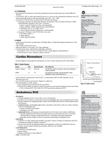
Table 4. Cardiac Enzymes
80 75 70 65 60 55 50 45 40 35 30 25 20 15 10 5
Troponin T (early reperfusion)
Troponin I
CPK/CPK-MB
Troponin T (permanent occlusion)
Enzyme
Troponin I, Troponin T CK-MB
Peak
1-2 d 1 d
Duration Elevated
Up to 2 wk 3 d
DDx of Elevation
MI, CHF, AFib, acute PE, myocarditis, chronic renal insufficiency, sepsis, hypovolemia
MI, myocarditis, pericarditis, muscular dystrophy, cardiac defibrillation, chronic renal insufficiency, etc.
• checktroponinIatpresentationand8hlater±creatinekinase-MB(CK-MB;dependsonlocal laboratory protocol)
• new CK-MB elevation can be used to diagnose re-infarction
• otherbiomarkersofcardiacdisease
■ ASTandLDHalsoincreasedinMI(lowspecificity)
■ BNPandNT-proBNP:secretedbyventriclesinresponsetoincreasedend-diastolicpressureandvolume
◆ DDx of elevated BNP: CHF, AFib, PE, COPD exacerbation, pulmonary HTN
Ambulatory ECG
• description
■ extendedambulatoryECGof24or48hor14or28dduration
■ providesaviewofonlytwoorthreeleadsofelectrocardiographicdataoveranextendedperiodoftime ■ permits evaluation of changing dynamic cardiac electrical phenomena that are often transient and of
brief duration
■ continuousloop:asmall,lightweight,batteryoperatedrecorderthatrecordstwoorthreechannels
of electrocardiographic data
◆ patient activated event markers ◆ minimum of 24-48 h
■ implantabledevice:subcutaneousmonitoringdeviceforthedetectionofcardiacarrhythmias ◆ typically implanted in the left pectoral region and stores events when the device is activated
automatically according to programmed criteria or manually with magnet application ◆ generally used for months to years of continuous monitoring for infrequent events
• indications
■ evaluation of cardiac rhythm abnormalities; typically to obtain correlation of symptoms with ECG:
“instantaneous symptom-rhythm correlation”
■ has also been used for assessing pacemaker and implantable cardioverter-defibrillator function,
evidence of myocardial ischemia, late potentials, and heart rate variability
• contraindications
■ no absolute contraindications
■ patient refusal
■ allergies (sensitivities to latex adhesive)
12 24
Time after acute myocardial infarction (h)
© Susan Park 2009
36 48 60
72 84
96 108 120 132 144 156 168 180 192
Figure 16. Cardiac enzymes
Use of B-Type Natriuretic Peptide in the Evaluation and Management of Acute Dyspnea (BASEL)
NEJM 2004;350;647-54
Study: Prospective, RCT.
Population: 452 patients (mean age 71 yr, 58% male) with acute dyspnea; patients with severe renal disease or cardiogenic shock were excluded. Intervention: Assessment including measurement of B-type natriuretic peptide or standard assessment. Outcome: Time to discharge and total cost of treatment.
Results: Median time to discharge was significantly shorter in the intervention group when compared with the control group (8.0 vs. 11.0 d, p=0.001). Total cost was also significantly lower in the intervention group ($5410 vs. $7264, p=0.006).
In addition, the measurement of B-type natriuretic peptide significantly reduced the need for admission to hospital and intensive care. The 30-d mortality rates were similar (10% vs. 12%, p=0.45). Conclusions: In patients with acute dyspnea, measurement of B-type natriuretic peptide improves clinical outcomes (need for hospitalization or intensive care) and reduces time to discharge and total cost of treatment.
• risks:noabsoluterisks
Enzyme levels (ng/mL)
