
Page 90 - TNFlipTest
P. 90
C22 Cardiology and Cardiac Surgery
Arrhythmias Toronto Notes 2019
• treatment:electricalcardioversion,IVprocainamide,orIVamiodarone
■ do not use drugs that slow AV node conduction (digoxin, β-blockers) as this may cause preferential
conduction through the bypass tract and precipitate VF ■ long-term: ablation of bypass tract if possible
AV Re-Entrant Tachycardia
• re-entrantloopviaaccessorypathwayandnormalconductionsystem
• initiatedbyaprematureatrialorventricularcomplex
• orthodromic AVRT: stimulus from a premature complex travels up the bypass tract (V to A) and
down the AV node (A to V) with narrow QRS complex (no delta wave because stimulus travels through
normal conduction system)
• comprises95%ofthereentranttachycardiasassociatedwithWPWsyndrome
• antidromicAVRT:morerarelythestimulusgoesuptheAVnode(VtoA)anddownthebypasstract
(A to V); wide and abnormal QRS as ventricular activation is only via the bypass tract • treatment
■ acute: similar to AVNRT except avoid long-acting AV nodal blockers (e.g. digoxin and verapamil) ■ long-term: for recurrent arrhythmias ablation of the bypass tract is recommended
■ drugs such as flecainide and procainamide can be used
Ventricular Tachyarrhythmias
Premature Ventricular Contraction (PVC) or Ventricular Premature Beat (VPB)
• QRSwidth>120msec,noprecedingPwave,bizarreQRSmorphology
• origin:LBBBmorphologyofVT=RVorigin;RBBBmorphologyofVT=LVorigin • PVCsmaybebenignbutareusuallysignificantinthefollowingsituations:
■ consecutive (≥3 = VT) or multiform (varied origin)
■ PVC falling on the T wave of the previous beat (“R on T phenomenon”): may precipitate ventricular
tachycardia or VF
• risk of sustained arrhythmia depends on the clinical situation (i.e. MI, HF), not the PVCs themselves
Accelerated Idioventricular Rhythm
• ectopicventricularrhythmwithrate50-100bpm
• morefrequentlyoccursinthepresenceofsinusbradycardiaandiseasilyoverdrivenbyafaster
supraventricular rhythm
• frequentlyoccursinpatientswithacuteMIorothertypesofheartdisease(cardiomyopathy,
hypertensive, valvular) but it does not affect prognosis and does not usually require treatment
Ventricular Tachycardia (VT)
• 3ormoreconsecutiveectopicventricularcomplexes
■ rate>100bpm(usually140-200)
■ ventricular flutter: if rate >200 bpm and complexes resemble a sinusoidal pattern
■ “sustained VT” if it lasts longer than 30 s
■ ECG characteristics: wide regular QRS tachycardia (QRS usually >140 msec)
■ AV dissociation; bizarre QRS pattern
■ also favour Dx of VT: left axis or right axis deviation, nonspecific intraventricular block pattern,
monophasic or biphasic QRS in V1 with RBBB, QRS concordance in V1-V6
■ occasionally during VT supraventricular impulses may be conducted to the ventricles generating QRS complexes with normal or aberrant supraventricular morphology (“ventricular capture”) or
summation pattern (“fusion complexes”)
• monomorphicVT
■ identical complexes with uniform morphology
■ more common than polymorphic VT
■ typically result from intraventricular re-entry circuit
■ potential causes: chronic infarct scarring, acute MI/ischemia, cardiomyopathies, myocarditis,
arrhythmogenic right ventricular dysplasia, idiopathic, drugs (e.g. cocaine), electrolyte disturbances
• polymorphicVT
■ complexes with constantly changing morphology, amplitude, and polarity
■ more frequently associated with hemodynamic instability due to faster rates (typically 200-250 bpm)
vs. monomorphic VT
■ potential causes: acute MI, severe or silent ischemia, and predisposing factors for QT prolongation
• treatment
■ sustained VT (>30 s) is an emergency, requiring immediate treatment
■ hemodynamic compromise: electrical cardioversion
■ no hemodynamic compromise: electrical cardioversion, lidocaine, amiodarone, type Ia agents
(procainamide, quinidine)
Figure 32. Ventricular tachycardia (monomorphic)
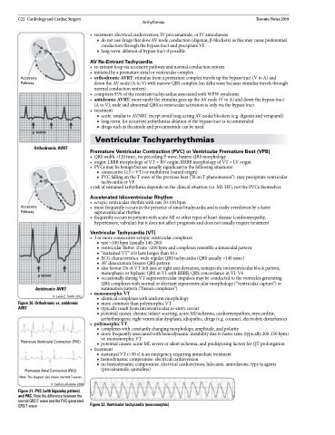
Accessory Pathway
Orthodromic AVRT
Accessory Pathway
Antidromic AVRT
© Laura E. Smith 2012
Figure 30. Orthodromic vs. antidromic AVRT
Premature Ventricular Contraction (PVC)
Premature Atrial Contraction (PAC) Note: This diagram also shows inverted T-waves
© Caitlin LaFlamme 2009
Figure 31. PVC (with bigeminy pattern) and PAC. Note the difference between the normal QRS/T wave and the PVC-generated QRS/T wave
