
Page 909 - TNFlipTest
P. 909
Toronto Notes 2019 The Uveal Tract Ophthalmology OP19 The Uveal Tract
• uvealtract(fromanteriortoposterior)=iris,ciliarybody,choroid
• vascularized,pigmentedmiddlelayeroftheeye,betweenthescleraandtheretina
Uveitis
• uvealinflammationwhichmayinvolveone,two,orallthreepartsofthetract
• idiopathicorassociatedwithautoimmune,infectious,granulomatous,andmalignantcauses
• shouldbemanagedbyanoptometristorophthalmologist
• anatomicallyclassifiedasanterioruveitis,intermediateuveitis,posterioruveitis,orpanuveitisbasedon
primary site of inflammation
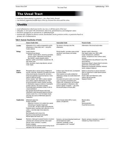
Table 8. Anatomic Classification of Uveitis
Location Etiology
Clinical Features
Anterior Uveitis (Iritis)
Inflammation of iris, usually accompanied by cyclitis (inflammation of ciliary body), both = iridocyclitis Usually unilateral
Usually idiopathic Connective tissue diseases:
HLA-B27: reactive arthritis, ankylosing spondylitis, psoriatic arthritis, inflammatory bowel disease Non-HLA-B27: juvenile idiopathic arthritis
Infectious: syphilis, Lyme disease, toxoplasmosis, TB, HSV, herpes zoster
Other: sarcoidosis, trauma, large abrasion, and post ocular surgery
Photophobia (due to reactive spasm of inflamed iris muscle), ocular pain, tenderness of the globe, brow ache (ciliary muscle spasm), decreased VA, lacrimation Ciliary flush (perilimbal conjunctival injection), miosis (spasm of sphincter muscle)
Anterior chamber “cells” (WBC in anterior chamber
due to anterior segment inflammation) and “flare” (protein precipitates in anterior chamber secondary
to inflammation), hypopyon (collection of neutrophilic exudates inferiorly in the anterior chamber)
Occasionally keratic precipitates (clumps of cells on corneal endothelium)
Iritis typically reduces IOP because ciliary body inflammation causes decreased aqueous production; however, severe iritis, or iritis from herpes simplex
and zoster may cause an inflammatory glaucoma (trabeculitis)
Inflammatory glaucoma Posterior synechiae
Adhesions of posterior iris to anterior lens capsule Indicated by an irregularly shaped pupil
If occurs 360°, can lead to angle closure glaucoma
Peripheral anterior synechiae (rare): adhesions of iris to cornea → secondary angle closure glaucoma Cataracts
Band keratopathy (with chronic iritis)
Superficial corneal calcification keratopathy Macular edema with chronic iritis
Mydriatics: dilate pupil to prevent formation of posterior synechiae and to decrease pain from ciliary spasm Steroids: topical, sub-tenon, or systemic
Systemic analgesia
If recurrent episodes, extensive medical workup may be indicated to rule out secondary causes
Intermediate Uveitis
The vitreous is the major site of the inflammation
Mostly idiopathic, secondary causes include sarcoidosis, Lyme disease, and multiple sclerosis
Insidious onset of blurred vision, accompanied by vitreous floaters
Initial symptoms are usually unilateral but inflammatory changes are usually bilateral and asymmetric
Associated with anterior uveitis, most severe cases of secondary intermediate uveitis Vitreous cells, condensations, and snowballs (vitreous aggregates of inflammatory cells) Posterior segment ‘snowbank’ = grey-white fibrovascular plaque at the pars plana
Cystoid macular edema (30% of cases), cataract, and glaucoma
Systemic or sub-tenon/intravitreal steroids and immunosuppressive agents
Vitrectomy, cryotherapy, or laser photocoagulation to the “snowbank”
Posterior Uveitis
Inflammation of the choroid and/or retina
Bacterial: syphilis, tuberculosis
Viral: herpes simplex virus, CMV in AIDS
Fungal: histoplasmosis, candidiasis
Parasitic: toxoplasmosis (most common cause), toxocara
Immunosuppression may predispose to any of the above infections
Autoimmune: Behçet’s disease (triad of oral ulcers, genital ulcers, and posterior uveitis)
Malignancies (masquerade syndrome): metastatic lesions, malignant melanoma
Painless as choroid has no sensory innervation Often no conjunctival or scleral injection present Decreased VA
Floaters (debris and inflammatory cells) Vitreous cells and opacities
Hypopyon formation
Macular edema
Vitritis Neovascularization Visual field loss/scotoma
Steroids: sub-tenon, intravitreal, or systemic if indicated (e.g. threat of vision loss)
Complications
Treatment
