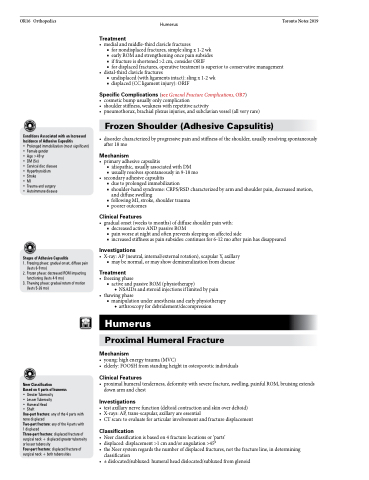
Page 950 - TNFlipTest
P. 950
 OR16 Orthopedics
Humerus
Toronto Notes 2019
   Conditions Associated with an Increased Incidence of Adhesive Capsulitis
• Prolonged immobilization (most significant) • Female gender
• Age >49 yr
• DM(5x)
• Cervical disc disease • Hyperthyroidism
• Stroke
• MI
• Trauma and surgery • Autoimmune disease
Stages of Adhesive Capsulitis
1. Freezing phase: gradual onset, diffuse pain (lasts 6-9 mo)
2. Frozen phase: decreased ROM impacting functioning (lasts 4-9 mo)
3. Thawing phase: gradual return of motion (lasts 5-26 mo)
Treatment
• medialandmiddle-thirdclaviclefractures
■ for nondisplaced fractures, simple sling x 1-2 wk
■ earlyROMandstrengtheningoncepainsubsides
■ if fracture is shortened >2 cm, consider ORIF
■ for displaced fractures, operative treatment is superior to conservative management
• distal-thirdclaviclefractures
■ undisplaced (with ligaments intact): sling x 1-2 wk ■ displaced (CC ligament injury): ORIF
Specific Complications (see General Fracture Complications, OR7)
• cosmeticbumpusuallyonlycomplication
• shoulderstiffness,weaknesswithrepetitiveactivity
• pneumothorax,brachialplexusinjuries,andsubclavianvessel(allveryrare)
Frozen Shoulder (Adhesive Capsulitis)
• disordercharacterizedbyprogressivepainandstiffnessoftheshoulder,usuallyresolvingspontaneously after 18 mo
Mechanism
• primary adhesive capsulitis
■ idiopathic, usually associated with DM
■ usually resolves spontaneously in 9-18 mo
• secondaryadhesivecapsulitis
■ due to prolonged immobilization
■ shoulder-hand syndrome: CRPS/RSD characterized by arm and shoulder pain, decreased motion,
and diffuse swelling
■ following MI, stroke, shoulder trauma
■ poorer outcomes
Clinical Features
• gradualonset(weekstomonths)ofdiffuseshoulderpainwith:
■ decreased active AND passive ROM
■ pain worse at night and often prevents sleeping on affected side
■ increased stiffness as pain subsides: continues for 6-12 mo after pain has disappeared
Investigations
• X-ray:AP(neutral,internal/externalrotation),scapularY,axillary ■ may be normal, or may show demineralization from disease
Treatment
• freezingphase
■ activeandpassiveROM(physiotherapy)
◆ NSAIDs and steroid injections if limited by pain • thawingphase
■ manipulation under anesthesia and early physiotherapy ◆ arthroscopy for debridement/decompression
Humerus
Proximal Humeral Fracture
Mechanism
• young:highenergytrauma(MVC)
• elderly:FOOSHfromstandingheightinosteoporoticindividuals
Clinical Features
• proximalhumeraltenderness,deformitywithseverefracture,swelling,painfulROM,bruisingextends down arm and chest
Investigations
• testaxillarynervefunction(deltoidcontractionandskinoverdeltoid)
• X-rays:AP,trans-scapular,axillaryareessential
• CTscan:toevaluateforarticularinvolvementandfracturedisplacement
Classification
• Neerclassificationisbasedon4fracturelocationsor‘parts’
• displaced: displacement >1 cm and/or angulation >45°
• the Neer system regards the number of displaced fractures, not the fracture line, in determining
classification
• ±dislocated/subluxed:humeralheaddislocated/subluxedfromglenoid
       Neer Classification
Based on 4 parts of humerus
• Greater Tuberosity
• Lesser Tuberosity
• Humeral Head
• Shaft
One-part fracture: any of the 4 parts with none displaced
Two-part fracture: any of the 4 parts with 1 displaced
Three-part fracture: displaced fracture of surgical neck + displaced greater tuberosity or lesser tuberosity
Four-part fracture: displaced fracture of surgical neck + both tuberosities


