
Page 952 - TNFlipTest
P. 952
OR18 Orthopedics
Elbow Toronto Notes 2019
Three Joints at the Elbow
• Humeroradial joint • Humeroulnar joint • Radioulnar joint
Normal carrying angle of elbow is ~10° of valgus
Humeral shaft
Radius
Elbow
Supracondylar Fracture
• subclassofdistalhumerusfracture:extra-articular,fractureproximaltocapitulumandtrochlea,usually transverse
• mostcommoninpediatricpopulation(peakage~7yrold),rarelyseeninadults
• AIN(mediannerve)injurycommonlyassociatedwithextensiontype
Mechanism
• >96%areextensioninjuriesviaFOOSH(e.g.falloffmonkeybars);<4%areflexioninjuries
Clinical Features
• pain,swelling,pointtenderness
• neurovascular injury: median and radial nerves, radial artery
Investigations
• X-ray:AP,lateralofelbow
■ disruption of anterior humeral line suggests supracondylar fracture
■ fat pad sign: a sign of effusion and can be indicative of occult fracture ■ assess NVS: median and radial nerves, radial artery
Treatment
• non-operative
■ nondisplaced: long arm plaster slab in 90° flexion x 3 wk
• operative
■ indications: displaced>50%, vascular injury, open fracture
■ requires percutaneous pinning followed by limb cast with elbow flexed <90° ■ in adults, ORIF is necessary
Specific Complications (see General Fracture Complications, OR7)
• stiffnessismostcommon
• brachialarteryinjury(kinkingcanoccurifdisplacedfracture),medianorulnarnerveinjury,
compartment syndrome (leads to Volkmann’s ischemic contracture), malalignment cubitus varus (distal fragment tilted into varus)
Radial Head Fracture
• acommonfractureoftheupperlimbinyoungadults
Mechanism
• FOOSHwithelbowextendedandforearmpronated
Clinical Features
• markedlocaltendernessonpalpationoverradialhead(lateralelbow)
• decreasedROMatelbow,±mechanicalblocktoforearmpronationandsupination • painonpronation/supination
Investigations
• X-ray:enlargedanteriorfatpad(“sailsign”)orthepresenceofaposteriorfatpadindicateseffusion which could occur with occult radial head fractures
Table 12. Classification and Treatment of Radial Head Fractures
Ulna
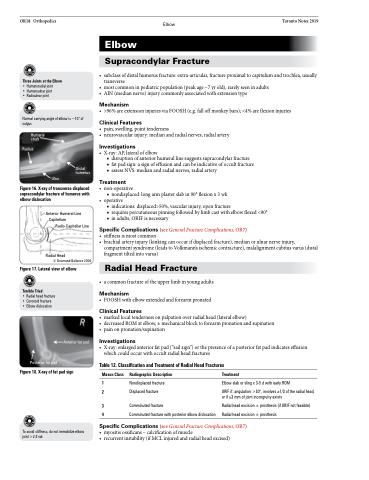
Figure 16. X-ray of transverse displaced supracondylar fracture of humerus with elbow dislocation
Anterior Humeral Line Capitellum
Radio-Capitellar Line
Radial Head
© Desmond Ballance 2006
Figure 17. Lateral view of elbow
Distal humerus
Terrible Triad
• Radial head fracture • Coronoid fracture
• Elbowdislocation
Posterior fat pad
Anterior fat pad
Figure 18. X-ray of fat pad sign
To avoid stiffness, do not immobilize elbow joint >2-3 wk
Mason Class
1 2
3 4
Radiographic Description
Nondisplaced fracture Displaced fracture
Comminuted fracture
Comminuted fracture with posterior elbow dislocation
Treatment
Elbow slab or sling x 3-5 d with early ROM
ORIF if: angulation >30°, involves ≥1/3 of the radial head,
or if ≥3 mm of joint incongruity exists
Radial head excision ± prosthesis (if ORIF not feasible) Radial head excision ± prosthesis
Specific Complications (see General Fracture Complications, OR7) • myositisossificans–calcificationofmuscle
• recurrentinstability(ifMCLinjuredandradialheadexcised)
