
Page 954 - TNFlipTest
P. 954
OR20 Orthopedics
Forearm
Toronto Notes 2019
Elbow Joint Injection
Inject at the centre of the triangle formed by the lateral epicondyle, radial head, and olecranon
Treatment
• non-operative(verygoodoutcomes)
■ rest, ice, NSAIDs
■ use brace/strap
■ physiotherapy, stretching, and strengthening ■ corticosteroid injection
• operative
■ indication: failed 6-12 mo conservative therapy
■ percutaneous or open release of common tendon from epicondyle
Forearm
Radius and Ulna Shaft Fractures
Mechanism
• high-energydirectorindirect(MVA,fallfromheight,sports)trauma • fracturesusuallyaccompaniedbydisplacementduetohighforce
Clinical Features
• deformity, pain, swelling
• lossoffunctioninhandandforearm
Investigations
• X-ray:APandlateralofforearm±obliqueofelbowandwrist • CTiffractureisclosetojoint
Treatment
• goalisanatomicreductionsinceimperfectalignmentsignificantlylimitsforearmpronationand supination
• ORIFwithplatesandscrews;closedreductionwithimmobilizationusuallyyieldspoorresultsfor displaced forearm fractures (except in children)
Specific Complications (see General Fracture Complications, OR7)
• softtissuecontractureresultinginlimitedforearmrotation–surgicalreleaseoftissuemaybewarranted
Monteggia Fracture
• fractureoftheproximalulnawithradialheaddislocationandproximalradioulnarjointinjury • morecommonandbetterprognosisinthepediatricagegroupwhencomparedtoadults
Mechanism
• directblowontheposterioraspectoftheforearm • hyperpronation
• fallonthehyperextendedelbow
Clinical Features
• pain,swelling,decreasedrotationofforearm±palpablelumpattheradialhead
• ulnaangledapexanteriorandradialheaddislocatedanteriorly(rarelythereversedeformityoccurs)
Investigations
• X-ray:AP,lateralelbow,wristandforearm
Treatment
• adults:ORIFofulnawithindirectradiusreductionin90%ofpatients(ORIFofradiusifunsuccessful)
• splintandearlypost-operativeROMifelbowcompletelystable,otherwiseimmobilizationinplaster
with elbow flexed for 6 wk
• pediatrics:attemptclosedreductionandimmobilizationinplasterwithelbowflexedforBadoType
I-III, surgery for Type IV
Specific Complications (see General Fracture Complications, OR7)
• PIN:mostcommonnerveinjury;observefor3moasmostresolvespontaneously • radialheadinstability/redislocation
• radioulnarsynostosis
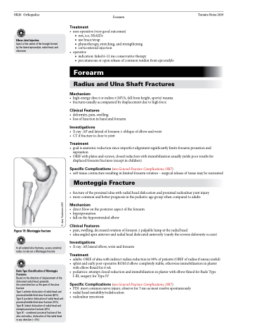
Figure 19. Monteggia fracture
In all isolated ulna fractures, assess proximal radius to rule out a Monteggia fracture
Bado Type Classification of Monteggia Fractures
Based on the direction of displacement of the dislocated radial head, generally
the same direction as the apex of the ulnar fracture
Type I: anterior dislocation of radial head and proximal/middle third ulnar fracture (60%) Type II: posterior dislocation of radial head and proximal/middle third ulnar fracture (15%) Type III: lateral dislocation of radial head and metaphyseal ulnar fracture (20%)
Type IV – combined: proximal fracture of the ulna and radius, dislocation of the radial head in any direction (<5%)
© Joey Trautmann 2007
