
Page 966 - TNFlipTest
P. 966
OR32 Orthopedics
Knee Toronto Notes 2019
135
4
2
6
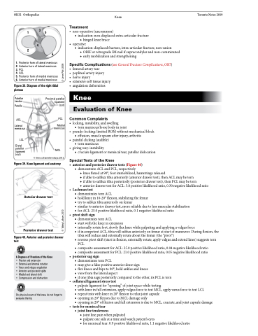
1. Posterior horn of lateral meniscus 2. Anterior horn of lateral meniscus 3. PCL
4. ACL
5. Posterior horn of medial meniscus 6. Anterior horn of medial meniscus
Figure 38. Diagram of the right tibial plateau
Treatment
• non-operative(uncommon)
■ indication: non-displaced extra-articular fracture
◆ hinged knee brace • operative
■ indication: displaced fracture, intra-articular fracture, non-union
◆ ORIF or retrograde IM nail if supracondylar and non-comminuted ◆ early mobilization and strengthening
Specific Complications (see General Fracture Complications, OR7) • femoralarterytear
• popliteal artery injury
• nerveinjury
• extensivesofttissueinjury • angulation deformities
Knee
Evaluation of Knee
Common Complaints
• locking, instability, and swelling
■ torn meniscus/loose body in joint
• pseudo-locking:limitedROMwithoutmechanicalblock ■ effusion, muscle spasm after injury, arthritis
• painful clicking (audible) ■ tornmeniscus
• giving way: instability
■ cruciate ligament or meniscal tear, patellar dislocation
Special Tests of the Knee
• anteriorandposteriordrawertests(Figure40) ■ demonstrate ACL and PCL, respectively
◆ knee flexed at 90°, foot immobilized, hamstrings released
◆ if able to sublux tibia anteriorly (anterior drawer test), then ACL may be torn
◆ if able to sublux tibia posteriorly (posterior drawer test), then PCL may be torn
◆ anterior drawer test for ACL: 3.8 positive likelihood ratio, 0.30 negative likelihood ratio
• Lachmantest
■ demonstrates torn ACL
■ hold knee in 10-20° flexion, stabilizing the femur
■ try to sublux tibia anteriorly on femur
■ similar to anterior drawer test, more reliable due to less muscular stabilization ■ for ACL: 25.0 positive likelihood ratio, 0.1 negative likelihood ratio
• pivotshiftsign
■ demonstrates torn ACL
■ start with the knee in extension
■ internally rotate foot, slowly flex knee while palpating and applying a valgus force
■ if incompetent ACL, tibia will sublux anteriorly on femur at start of maneuver. During flexion, the
tibia will reduce and externally rotate about the femur (the “pivot”)
■ reverse pivot shift (start in flexion, externally rotate, apply valgus and extend knee) suggests torn
PCL
■ composite assessment for ACL: 25.0 positive likelihood ratio, 0.04 negative likelihood ratio ■ composite assessment for PCL: 21.0 positive likelihood ratio, 0.05 negative likelihood ratio
• posterior sag sign
■ demonstrates torn PCL
■ may give a false positive anterior draw sign
■ flex knees and hips to 90°, hold ankles and knees
■ viewfromthelateralaspect
■ if one tibia sags posteriorly compared to the other, its PCL is torn
• collateralligamentstresstest
■ palpate ligament for “opening” of joint space while testing
■ with knee in full extension, apply valgus force to test MCL, apply varus force to test LCL
■ repeat tests with knee in 20° flexion to relax joint capsule
■ opening in 20° flexion due to MCL damage only
■ opening in 20° of flexion and full extension is due to MCL, cruciate, and joint capsule damage
• testsformeniscaltear
■ jointlinetenderness
◆ joint line pain when palpated
◆ palpate one side at a time and watch patient’s eyes
◆ for meniscal tear: 0.9 positive likelihood ratio, 1.1 negative likelihood ratio
Patellar tendon
Patella
ACL
Proximal patellar ligament (cut)
PCL
Lateral Medial meniscus meniscus
LCL
Distal patellar ligament (cut)
MCL
© Inessa Stanishevskaya 2012
Figure 39. Knee ligament and anatomy
Anterior drawer test
Posterior drawer test
Figure 40. Anterior and posterior drawer test
6 Degrees of Freedom of the Knee
• Flexion and extension
• External and internal rotation • Varus and valgus angulation • Anterior and posterior glide • Medial and lateral shift
• Compression and distraction
On physical exam of the knee, do not forget to evaluate the hip
© Tabby Lulham 2010 © Jenn Platt 2004
