
Page 967 - TNFlipTest
P. 967
Toronto Notes 2019 Knee
■ crouchcompressiontest
◆ joint line pain when squatting (anterior pain suggests patellofemoral pathology)
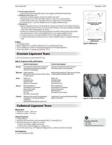
■ McMurray’stest(Figure41)
◆ with knee in flexion, palpate joint line for painful “pop/click”
◆ lateral meniscus tear exam: internally rotate foot, varus stress, and extend knee
◆ medial meniscus tear exam: externally rotate foot, valgus stress, and extend knee ◆ for meniscal tear: 1.3 positive likelihood ratio, 0.8 negative likelihood ratio
■ Thessaly test
◆ patient stands flat footed on one leg while the examiner provides his or her hands for balance.
The patient then flexes the knee to 20° and rotates the femur on the tibia medially and laterally
three times while maintaining the 20° flexion
◆ positive for a meniscal tear if the patient experiences medial or lateral joint line discomfort
◆ for medial meniscus: 29.67 positive likelihood ratio, 0.11 negative likelihood ratio
◆ for lateral meniscus: 23.0 positive likelihood ratio, 0.083 negative likelihood ratio
◆ composite assessment for meniscal tears: 2.7 positive likelihood ratio, 0.4 negative likelihood
ratio
X-Rays
• APstanding,lateral
• skyline:tangentialviewwithkneesflexedat45°toseepatellofemoraljoint
• 3-footstandingview:usefulinevaluatingleglengthandvarus/valgusalignment • OttawaKneeRules(seeEmergencyMedicine,ER16)
Orthopedics OR33
Examination for medial meniscal tear
Examination for lateral meniscal tear
Figure 41. McMurray test
Cruciate Ligament Tears
• ACLtearmuchmorecommonthanPCLtear
Table 22. Comparison of ACL and PCL Injuries
Anatomy Mechanism History
Physical
Treatment
Anterior Cruciate Ligament
From medial wall of lateral femoral condyle to the anteromedial and posterolateral intercondyloid eminence of the tibial plateau
Sudden deceleration
Hyperextension and internal rotation of tibia on femur (i.e. “plant and turn”)
Audible “pop”
Immediate swelling
Knee “giving way”
Inability to continue activity
Effusion (hemarthrosis) Posterolateral joint line tenderness Positive anterior drawer
Positive Lachmann
Pivot shift
Test for MCL, meniscal injuries
Stable knee with minimal functional impairment: immobilization 2-4 wk with early ROM and strengthening
High demand lifestyle: ligament reconstruction
Posterior Cruciate Ligament
Lateral wall of medial femoral condyle to posterior intercondyloid eminence of the tibial plateau
Sudden posterior displacement of tibia when knee is flexed or hyperextended (e.g. dashboard MVC injury)
Audible “pop” Immediate swelling Pain with push off Cannot descend stairs
Effusion (hemarthrosis) Anteromedial joint line tenderness Positive posterior drawer
Reverse pivot shift
Other ligamentous, bony injuries
Unstable knee or young person/high-demand lifestyle: ligament reconstruction
ACL PCL
Figure 42. T1 MRI of torn ACL and PCL
O’Donoghue’s Unhappy Triad
• ACL rupture
• MCL rupture
• Meniscal damage (medial and/or lateral)
Collateral Ligament Tears
Mechanism
• valgusforcetoknee=MCLtear • varusforcetoknee=LCLtear
Clinical Features
• swelling/effusion
• tendernessaboveandbelowjointlinemedially(MCL)orlaterally(LCL) • joint laxity with varus or valgus force to knee
■ laxity with endpoint suggests partial tear
■ laxity with no endpoint suggests a complete tear
• testforotherinjuries(e.g.O’Donoghue’sunhappytriad),commonperonealnerveinjury
Investigations
• x-ray:APandlateral;MRI
© Tabby Lulham 2010
