
Page 978 - TNFlipTest
P. 978
OR44 Orthopedics
Pediatric Orthopedics Toronto Notes 2019 ■ mild/moderate slip: stabilize physis with pins in current position
2
1
Complications
• AVN(roughlyhalfofunstablehips),chondrolysis(lossofarticularcartilage,resultinginnarrowingof joint space), pin penetration, premature OA, loss of ROM
Developmental Dysplasia of the Hip
Definition
• abnormaldevelopmentofhip,resultingindysplasiaandsubluxation/dislocationofhip • mostcommonorthopedicdisorderinnewborns
Etiology
• duetoligamentouslaxity,muscularunderdevelopment,andabnormalshallowslopeofacetabularroof • spectrumofconditions
■ dislocated femoral head completely out of acetabulum
■ dislocatable head in socket
■ head subluxates out of joint when provoked
■ dysplastic acetabulum, more shallow and more vertical than normal
Physical Exam
• diagnosisisclinical
■ limited abduction of the flexed hip (<60°)
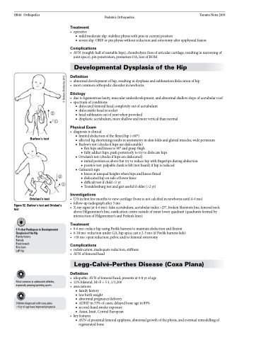
■ affected leg shortening results in asymmetry in skin folds and gluteal muscles, wide perineum ■ Barlow’s test (checks if hips are dislocatable)
Treatment
• operative
■ severe slip: ORIF or pin physis without reduction and osteotomy after epiphyseal fusion
Barlow’s test
2
1
◆ flex hips and knees to 90° and grasp thigh
◆ fully adduct hips, push posteriorly to try to dislocate hips ■ Ortolani’s test (checks if hips are dislocated)
◆ initial position as above but try to reduce hip with fingertips during abduction
◆ positive test: palpable clunk is felt (not heard) if hip is reduced ■ Galeazzi’s sign
◆ knees at unequal heights when hips and knees flexed ◆ dislocated hip on side of lower knee
◆ difficult test if child <1 yr
◆ Trendelenburg test and gait useful if older (>2 yr)
Ortolani’s test
Investigations
• U/S in first few months to view cartilage (bone is not calcified in newborns until 4-6 mo)
• follow-upradiographafter3mo
• X-raysigns(at4-6mo):falseacetabulum,acetabularindex>25°,brokenShenton’sline,femoralneck
above Hilgenreiner’s line, ossification centre outside of inner lower quadrant (quadrants formed by intersection of Hilgenreiner’s and Perkin’s lines)
Treatment
• 0-6mo:reducehipusingPavlikharnesstomaintainabductionandflexion
• 6-18mo:reductionunderGA,hipspicacastx2-3mo(ifPavlikharnessfails) • >18mo:openreduction;pelvicand/orfemoralosteotomy
Complications
• redislocation,inadequatereduction,stiffness • AVNoffemoralhead
Legg-Calvé-Perthes Disease (Coxa Plana)
Definition
• idiopathicAVNoffemoralhead,presentsat4-8yrofage • 12%bilateral,M>F=5:1,1/1,200
• associations
■ family history
■ lowbirthweight
■ abnormal pregnancy/delivery
■ ADHD in 33% of cases, delayed bone age in 89% ■ second-hand smoke exposure
■ Asian, Inuit, Central European
• keyfeatures
■ AVN of proximal femoral epiphysis, abnormal growth of the physis, and eventual remodelling of
Figure 52. Barlow’s test and Ortolani’s test
5 Fs that Predispose to Developmental Dysplasia of the Hip
Family history
Female
Frank breech First born LeFt hip
Most common in adolescent athletes, especially jumping/sprinting sports
Children diagnosed with coxa plana <6 yr of age have improved prognosis
regenerated bone
© Barbara Brehovsky 2012
