
Page 979 - TNFlipTest
P. 979
Toronto Notes 2019 Pediatric Orthopedics
Clinical Features
• childwithantalgicorTrendelenburggait±pain
• intermittentknee,hip,groin,orthighpain
• flexioncontracture(stiffhip):decreasedinternalrotationandabductionofhip • limblengthdiscrepancy(late)
Investigations
• X-ray:APpelvis,frogleglaterals
• maybenegativeearly(ifhighindexofsuspicion,movetobonescanorMRI) • eventually,characteristiccollapseoffemoralhead(diagnostic)
Treatment
• goalistopreserveROMandkeepfemoralheadcontainedinacetabulum • non-operative
■ physiotherapy:ROMexercises
■ brace in flexion and abduction x 2-3 yr (controversial) • operative
■ femoral or pelvic osteotomy (>8 yr of age or severe)
◆ prognosis better in males, <6 yr, <50% of femoral head involved, abduction >30°
• 60%ofinvolvedhipsdonotrequireoperativeintervention • naturalhistoryisearlyonsetOAanddecreasedROM
Osgood-Schlatter Disease
Definition
• inflammationofpatellarligamentatinsertionpointontibialtuberosity • M>F
• ageofonset:boys12-15yr;girls8-12yr
Mechanism
• repetitivetensilestressoninsertionofpatellartendonoverthetibialtuberositycausesminoravulsionat the site and subsequent inflammatory reaction (tibial tubercle apophysitis)
Clinical Features
• tenderlumpovertibialtuberosity
• painonresistedlegextension
• anteriorkneepainexacerbatedbyjumpingorkneeling,relievedbyrest
Investigations
• X-raylateralknee:fragmentationofthetibialtubercle,±ossiclesinpatellartendon
Treatment
• benign,self-limitedcondition,doesnotresolveuntilgrowthhalts • non-operative(majority)
■ may restrict activities such as basketball or cycling
■ NSAIDs, rest, flexibility, isometric strengthening exercises
■ casting if symptoms do not resolve with conservative management
• operative:ossicleexcisioninrefractorycases(patientisskeletallymaturewithpersistentsymptoms)
Congenital Talipes Equinovarus (Club Foot)
Definition
• congenital foot deformity
• musclecontracturesresultinginCAVEdeformity
• bonydeformity:talarneckmedialandplantardeviated;varuscalcaneusandrotatedmediallyaround
talus; navicular and cuboid medially displaced
Etiology
• intrinsiccauses(neurologic,muscular,orconnectivetissuediseases)vs.extrinsic(intrauterinegrowth restriction); may be idiopathic, neurogenic, or syndrome-associated
• fixeddeformity
• 1-2/1,000 newborns, 50% bilateral, occurrence M>F, severity F>M
Physical Exam
• examinehipsforassociatedDDH
• examinekneesfordeformity
• examinebackfordysraphism(unfusedvertebralbodies)
Orthopedics OR45
P A
S
P
AH
S
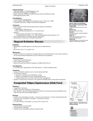
Figure 53. Pelvic x-ray and reference lines and angles for assessment of DDH Triradiate Cartilage
y-shaped epiphyseal plate at junction of ilium, ischium and pubis
Hilgenreiner’s Line
Line running between triradiate cartilages
Perkin’s Line
Line through lateral margin of acetabulum, perpendicular to Hilgenreiner’s Line
Shenton’s Line
Arced line along inferior border of femoral neck and superior margin of obturator foramen
Acetabular Index
Angle between Hilgenreiner’s Line and line from triradiate cartilage to point on lateral margin of acetabulum
Plantar flexion of ankle joint
Talus in equinus and varus
Forefoot bones in varus
Inversion of calcaneus
Figure 54. Club foot - depicting the gross and bony deformity
CAVE deformity
• Midfoot Cavus
• Forefoot Adductus • Hindfoot Varus
• Hindfoot Equinus
© Emilie McMahon 2005
