
Page 981 - TNFlipTest
P. 981
Toronto Notes 2019 Bone Tumours Table 25. Distinguishing Benign from Malignant Bone Lesions on X-Ray
Orthopedics OR47
Benign
No periosteal reaction
Thick endosteal reaction
Well developed bone formation Intraosseous and even calcification
Malignant
Acute periosteal reaction
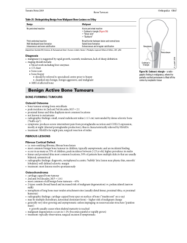
• Codman’s triangle (Figure 56) • “Onion skin”
• “Sunburst”
Broad border between lesion and normal bone Varied bone formation
Extraosseous and irregular calcification
Neoplasm
Codman’s triangle
Adapted from: Buckholtz RW, Heckman JD. Rockwood and Green’s Fractures in Adults. Volume 1. Philadephia: Lippincott Williams & Wilkins, 2001. p558
Diagnosis
• malignancyissuggestedbyrapidgrowth,warmth,tenderness,lackofsharpdefinition • stagingshouldinclude:
■ blood work including liver enzymes ■ CTchest
■ bone scan
■ bone biopsy
◆ should be referred to specialized centre prior to biopsy
◆ classified into benign, benign aggressive, and malignant ■ MRI of affected bone
Benign Active Bone Tumours
BONE-FORMING TUMOURS
Osteoid Osteoma
• bonetumourarisingfromosteoblasts
• peakincidencein2ndand3rddecades,M:F=2:1
• proximalfemurandtibiadiaphysismostcommonlocations
• notknowntometastasize
• radiographicfindings:small,roundradiolucentnidus(<1.5cm)surroundedbydensescleroticbone
(“bull’s-eye”)
• symptoms:producessevereintermittentpainfromprostaglandinsecretionandCOX1/2expression,
mostly at night (diurnal prostaglandin production), thus is characteristically relieved by NSAIDs • treatment:NSAIDsfornightpain;surgicalresectionofnidus
FIBROUS LESIONS
Fibrous Cortical Defect
• i.e.non-ossifyingfibroma,fibrousbonelesion
• mostcommonbenignbonetumourinchildren,typicallyasymptomaticandanincidentalfinding
• occurinasmanyas35%ofchildren,peakincidencebetween2-25yrold,higherprevalenceinmales
• femurandproximaltibiamostcommonlocations,50%ofpatientshavemultipledefectsthatareusually
bilateral, symmetrical
• radiographicfindings:diagnostic,metaphysealeccentric‘bubbly’lyticlesionnearphysis;thin,smooth/
lobulated, well-defined sclerotic margin
• treatment:mostlesionsresolvespontaneously
Osteochondroma
• cartilagecappedbonytumour
• 2ndand3rddecades,M:F=1.8:1
• mostcommonofallbenignbonetumours–45%
• 2types:sessile(broadbasedandincreasedriskofmalignantdegeneration)vs.pedunculated(narrow
stalk)
• metaphysisoflongboneneartendonattachmentsites(usuallydistalfemur,proximaltibia,orproximal
humerus)
• radiographicfindings:cartilage-cappedbonyspuronsurfaceofbone(“mushroom”onx-ray)
• maybemultiple(hereditary,autosomaldominantform)–higherriskofmalignantchange
• generallyveryslowgrowingandasymptomaticunlessimpingingonneurovascularstructure(‘painless
mass’)
■ growth usually ceases when skeletal maturity is reached
• malignantdegenerationoccursin1-2%(becomespainfulorrapidlygrows) • treatment:typicallyobservation;surgicalexcisionifsymptomatic
Periosteum
Figure 56. Codman’s triangle – a radio- graphic finding in malignancy, where the partially ossified periosteum is lifted off the cortex by neoplastic tissue
© j.a.platt 2005
