
Page 982 - TNFlipTest
P. 982
OR48 Orthopedics
Bone Tumours Toronto Notes 2019
Figure 57. T1MRI of femoral enchondroma
Figure 58. X-ray of aneurysmal bone cyst
Note the aggressive destruction of bone
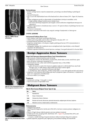
Figure 59. X-ray of osteosarcoma of distal femur
Enchondroma
• hyalinecartilagetumour;majorityasymptomatic,presentingasincidentalfindingorpathological fracture
• 2ndand3rddecades
• 60%occurinthesmalltubularbonesofthehandandfoot;othersinfemur(20%-Figure56),humerus,
ribs
• benigncartilaginousgrowth,anabnormalityofchondroblasts,developsinmedullarycavity
■ single/multiple enlarged rarefied areas in tubular bones
■ lytic lesion with sharp margination and irregular central calcification (stippled/punctate/popcorn
appearance)
• malignantdegenerationtochondrosarcomaoccursin1-2%(paininabsenceofpathologicfractureisan
important clue)
• notknowntometastasize
• treatment:observationwithserialx-rays;surgicalcurettageifsymptomaticorlesiongrows
CYSTIC LESIONS
Unicameral/Solitary Bone Cyst
• mostcommoncysticlesion;serousfluid-filledlesion
• childrenandyoungadults,peakincidenceduringfirst2decades,M:F=2:1
• proximalhumerusandfemurmostcommon
• symptoms:asymptomatic,orlocalpain;completepathologicalfracture(50%ofpresentations)or
incidental detection
• radiographicfindings:lytictranslucentareaonmetaphysealsideofgrowthplate,cortexthinned/
expanded; well-defined lesion
• treatment:aspirationfollowedbysteroidinjection;curettage±bonegraftindicatedifre-fracturelikely
Benign Aggressive Bone Tumours
Giant Cell Tumours/Aneurysmal Bone Cyst/Osteoblastoma
• affectspatientsofskeletalmaturity,peak3rddecade
• osteoblastoma:foundinthedistalfemur,proximaltibia,distalradius,sacrum,tarsalbones,spine • giantcelltumour:pulmonarymetastasesin3%
• aneurysmalbonecysts:eithersolidwithfibrous/granulartissue,orblood-filled
• radiographicfindings
■ giant cell tumour: eccentric lytic lesions in epiphyses adjacent to subchondral bone; may break through cortex; T2 MRI enhances fluid within lesion (hyper-intense signal)
■ aneurysmal bone cyst: expanded with honeycomb shape
■ osteoblastoma: often nonspecific; calcified central nidus (>2 cm) with radiolucent halo and sclerosis • symptoms:localtendernessandswelling,painmaybeprogressive(giantcelltumours),±symptomsof
nerve root compression (osteoblastoma) • 15%recurwithin2yrofsurgery
Treatment
• intralesionalcurettage+bonegraftorcement • widelocalexcisionofexpendablebones
Malignant Bone Tumours
Table 26. Most Common Malignant Tumour Types for Age
Age Tumour
<1 Neuroblastoma
1-10 Ewing’s of tubular bones
10-30 Osteosarcoma, Ewing’s of flat bones
30-40 Reticulum cell sarcoma, fibrosarcoma, periosteal osteosarcoma, malignant giant cell tumour, lymphoma >40 Metastatic carcinoma, multiple myeloma, chondrosarcoma
Osteosarcoma
• malignantbonetumour
• mostfrequentlydiagnosedin2nddecadeoflife(60%),2ndmostcommonprimarymalignancyin
adults
• historyofPaget’sdisease(elderlypatients),previousradiationtreatment
• predilectionforsitesofrapidgrowth:distalfemur(45%-Figure59),proximaltibia(20%),and
proximal humerus (15%)
■ invasive, variable histology; frequent metastases without treatment (lung most common)
• painful symptoms: progressive pain, night pain, poorly defined swelling, decreased ROM
