
Page 991 - TNFlipTest
P. 991
Toronto Notes 2019 Basic Anatomy Review Anatomical Triangles of the Neck
Anterior triangle
• boundedbyanteriorborderofSCM,midlineofneck,andlowerborderofmandible • dividedinto:
■ submentaltriangle:boundedbybothanteriorbellyofdigastricandhyoidbone
■ digastrictriangle:boundedbyanteriorandposteriorbelliesofdigastricandinferiorborderof
mandible
■ carotidtriangle:boundedbySCM,anteriorbellyofomohyoid,andposteriorbellyofdigastric
◆ contains: tail of parotid, submandibular gland, hypoglossal nerve, carotid bifurcation, and lymph nodes
Otolaryngology OT5
Paired Parasympathetic Ganglia of the Head and Neck
• Ciliary: pupillary constriction
• Pterygopalatine: lacrimal gland, nasal
mucosa
• Submandibular: submandibular, sublingual
glands
• Otic: parotid gland
Function of Facial Nerve
“Ears, Tears, Face, Taste”
Ears: stapedius muscle
Tears: lacrimation (lacrimal gland) and salivation (parotid)
Face: muscles of facial expression Taste: sensory anterior 2/3 of tongue (via chorda tympani)
• Left-sided enlargement of a supraclavicular node (Virchow’s node) may indicate an abdominal malignancy
• Right-sided enlargement may indicate malignancy of the mediastinum, lungs, or esophagus
• Occipital and/or posterior auricular node enlargement may indicate rubella
4 Strap Muscles of the Neck
• Thyrohyoid • Omohyoid
• Sternohyoid • Sternothyroid
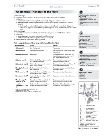
STA TC CCA
IJV CC
SPG * TG ITA IPG
Posterior triangle
• boundedbyposteriorborderofSCM,anteriorborderoftrapezius,andmiddlethirdofclavicle • dividedinto:
■ occipital triangle: superior to posterior belly of the omohyoid
■ subclaviantriangle:inferiortoposteriorbellyofomohyoid • contains:spinalaccessorynerveandlymphnodes
Table 1. Lymphatic Drainage of Nodal Groups and Anatomical Triangles of Neck
Nodal Group/Level
1. Suboccipital (S)
2. Retroauricular (R)
3. Parotid-preauricular (P)
4. Submental (Level IA)
5. Submandibular (Level IB)
6. Upper jugular (Levels IIA and IIB)
7. Middle jugular (Level III)
8. Lower jugular* (Level IV)
9. Posterior triangle** (Levels VA and VB)
10. Anterior compartment*** (Level VI)
Location
Base of skull, posterior Superficial to mastoid process
Anterior to ear
Anterior bellies (midline) of digastric muscles, tip of mandible, and hyoid bone
Anterior belly of digastric muscle, stylohyoid muscle, body of mandible
Skull base to inferior border of hyoid bone along SCM muscle
Inferior border of hyoid bone to inferior border of cricoid cartilage along SCM muscle
Inferior border of cricoid cartilage to clavicle along SCM muscle
Posterior border of SCM, anterior border of trapezius, from skull base to clavicle
Hyoid bone (midline) to suprasternal notch between the common carotid arteries
Drainage
Posterior scalp
Scalp, temporal region, external auditory meatus, posterior pinna
External auditory meatus, anterior pinna, soft tissue of frontal and temporal regions, root of nose, eyelids, palpebral conjunctiva
Floor of mouth, anterior tongue, anterior mandibular alveolar ridge, lower lip
Oral cavity, anterior nasal cavity, soft tissues of the mid-face, submandibular gland
Oral cavity, nasal cavity, naso/oro/hypopharynx, larynx, parotid glands
Oral cavity, naso/oro/hypopharynx, larynx
Hypopharynx, thyroid, cervical esophagus, larynx
Nasopharynx and oropharynx, cutaneous structures of the posterior scalp and neck
Thyroid gland, glottic and subglottic larynx, apex of piriform sinus, cervical esophagus
*Virchow node: left lower jugular (level IV) supraclavicular node **Includes some supraclavicular nodes
***Includes pretracheal, precricoid, paratracheal, and perithyroidal nodes
RRLN
VN (CN X)
LRLN
– Superior thyroid artery – Common carotid artery – Internal jugular vein
– Inferior thyroid artery
STA
CCA
IJV
ITA
RRLN –Rightrecurrentlaryngealnerve
TC
CC
SPG
TG
IPG
VN (CN X) LRLN
– Thyroid cartilage
– Cricoid cartilage
– Superior parathyroid gland
– Thyroid gland
– Inferior parathyroid gland
– Vagus nerve (CN X)
– Left recurrent laryngeal nerve
*Thyroidea ima artery: present in 3% of population, arises from aortic arch or innominate artery
Figure 10. Anatomy of the thyroid gland
© Erin Kenzie 2104 after Marisa Bonofiglio 2003
