
Page 989 - TNFlipTest
P. 989
Toronto Notes 2019
Basic Anatomy Review
Otolaryngology OT3
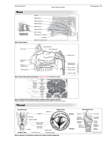
Nose
Speculum View of Right Nostril
Figure 3. Nasal anatomy
Frontal sinus
Kiesselbach’s plexus
Branch of superior labial a.
Greater palatine a.
Adenoid
Sphenoid sinus
Superior turbinate
Middle turbinate
Middle meatus
Inferior turbinate
Inferior meatus
Palatine process of maxilla
Soft palate Opening for Eustachian tube
Anterior ethmoid a.
Posterior ethmoid a.
Figure 4. Nasal septum and its arterial supply (see Epistaxis, OT26 for detailed blood supply) Frontal
sinus
Orbit
Ethmoid sinus
Lamina papyracea
Osteomeatal complex
Maxillary sinus
Nasal cavity
Teeth
Figure 5. Anatomy of the four paranasal sinuses: maxillary, ethmoid, sphenoid, and frontal
Reprinted from: Dhillon RS, East CA. Ear, Nose and Throat and Head and Neck Surgery, 2nd ed. Copyright 1999, with permission from Elsevier
Throat
Coronal Section
Superior View
Mid-Saggital View
Trachea
Pyriform fossa Arytenoid cartilage
Vestibule Thyroid cartilage
Vestibular ligaments (false cords)
Arytenoid cartilage
Vocal ligaments (true cords)
Epiglottis
Vestibular folds (false cords)
Vocal folds (true cords)
Anterior
Posterior
Epiglottis
Hyoid bone
Thyrohyoid membrane
Thyroid cartilage
Median cricothyroid ligament
Cricoid cartilage
Posterior View
Figure 6. Anatomy of a normal larynx; superior view of larynx on indirect laryngoscopy
Valeculla
Sphenoid sinus
Septal branch of sphenopalatine a.
Internal carotid a. External carotid a.
Common carotid a.
© Barbara Brehovsky 2012
© Jason Raine 2003
© Natalie Cormier 2015
© Glen Oomen 2002
