
Page 988 - TNFlipTest
P. 988
OT2 Otolaryngology Acronyms
ABR auditory brainstem response AC air conduction
AOM acute otitis media
BAHA bone anchored hearing aid BC bone conduction
C&S culture and sensitivity CHL conductive hearing loss CMV cytomegalovirus
CPA cerebellopontine angle DM diabetes mellitus
EAC external auditory canal
Acronyms
Toronto Notes 2019
Basic Anatomy Review
Ear
EBV Epstein-Barr virus
FAP familial adenomatous polyposis FESS functional endoscopic sinus surgery FNA fine needle aspiration
GERD gastroesophageal reflux disease GPA granulomatosis with polyangiitis H&N head and neck
HL hearing loss
HPV human papillomavirus
HSV herpes simplex virus
INCS intranasal corticosteroids
MEE middle ear effusion
MEI middle ear inflammation
MS multiple sclerosis
N/V nausea/vomiting
OE otitis externa
OME otitis media with effusion OSA obstructive sleep apnea
PMN polymorphonuclear leukocytes RA rheumatoid arthritis
RSV respiratory syncytial virus SCC squamous cell carcinoma
External Middle
Temporalis fascia Auditory ossicles and muscle
SCM sternocleidomastoid
SNHL sensorineural hearing loss
SRT speech reception threshold TEF tracheoesophageal fistula
TM tympanic membrane
TMJ temporomandibular joint TMP-SMX trimethoprim/sulfamethoxazole TNM tumour, node, metastases URTI upper respiratory tract infection
Helix Antihelix Helical crus
Malleus Incus Stapes
Inner
Semicircular canals
Vestibular nerve
Cochlear nerve
Triangular fossa
Scapha
Antitragus
© Aarti Inamdar
Tragus
Lobule
Facial nerve (CN VII) Cochlea
Eustachian tube
© Susan Park 2009
Tensor tympani tendon
Tensor tympani muscle
Tympanic plexus (branch of CN IX)
Hypotympanum Annulus
© Diana Dai 2006
Vestibulocochlear nerve (CN VIII)
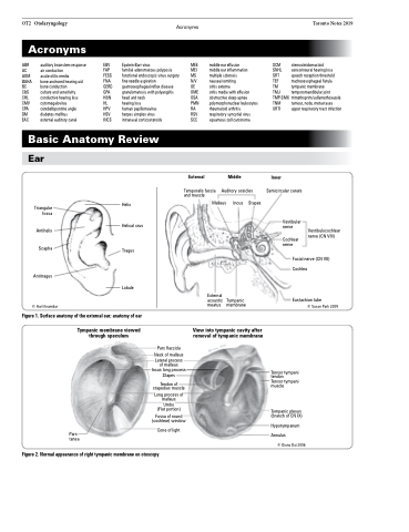
Figure 1. Surface anatomy of the external ear; anatomy of ear
Tympanic membrane viewed through speculum
View into tympanic cavity after removal of tympanic membrane
External acoustic meatus
Tympanic membrane
Pars tensa
Pars flaccida
Neck of malleus
Lateral process of malleus Incus long process Stapes
Tendon of stapedius muscle
Long process of malleus Umbo (Flat portion)
Fossa of round (cochlear) window
Cone of light
Figure 2. Normal appearance of right tympanic membrane on otoscopy
