
Page 1052 - TNFlipTest
P. 1052
P18 Pediatrics
Cardiology Toronto Notes 2019
■ clinical presentation
◆ history: delayed growth, decreased exercise tolerance, recurrent URTIs or “asthma” episodes ◆ physical exam: holosystolic murmur at LLSB, mid-diastolic rumble at apex, size of VSD is
inversely related to intensity of murmur ■ investigations
◆ ECG: LVH, LAH, RVH
◆ CXR: increased pulmonary vasculature, cardiomegaly, CHF ◆ Echo: diagnostic
■ management:treatmentofCHFandsurgicalclosureby1yrold
Patent Ductus Arteriosus
• patentvesselbetweendescendingaortaandleftpulmonaryartery(normally,functionalclosurewithin first 15 h of life, anatomical closure within first days of life)
• epidemiology
■ 5-10% of all congenital heart defects
■ delayed closure of ductus is common in premature infants (1/3 of infants <1,750 g); this is different
from PDA in term infants
• naturalhistory:spontaneousclosurecommoninprematureinfants,lesscommoninterminfants
• clinicalpresentation
■ history: asymptomatic, or have apneic or bradycardic spells, poor feeding, accessory muscle use, CHF ■ physical exam: tachycardia, bounding pulses, hyperactive precordium, wide pulse pressure,
continuous “machinery” murmur best heard at left infraclavicular area
• investigations
■ ECG: may show left atrial enlargement, LVH, RVH
■ ECHOisdiagnostic
■ CXR: normal to mildly enlarged heart, increased pulmonary vasculature, prominent pulmonary
artery
• management
■ indomethacin (Indocid®): antagonizes prostaglandin E2, which maintains ductus arteriosus patency; only effective in premature infants
■ catheterorsurgicalclosureifPDAcausesrespiratorycompromise,FTT,orpersistsbeyond3rdmooflife
2 . OBSTRUCTIVE LESIONS
• presentwithdecreasedurineoutput,pallor,coolextremitiesandpoorpulses,shock,orsuddencollapse
Coarctation of the Aorta
• definition:narrowingofaorta(almostalwaysattheleveloftheductusarteriosus)
• epidemiology:commonlyassociatedwithbicuspidaorticvalve(50%);Turnersyndrome(35%) • clinicalpresentation
■ history: often asymptomatic ■ physical exam
◆ blood pressure discrepancy between upper and lower extremities (increased suspicion/severity if >20 mmHg difference)
◆ diminished or delayed femoral pulses relative to brachial (i.e. brachial-femoral delay)
◆ possible systolic murmur with late peak at apex, left axilla, and left back
◆ if severe, presents with shock in the neonatal period when the ductus arteriosis closes
• investigations:ECGshowsRVHearlyininfancy,LVHlaterinchildhood;EchoorMRIfordiagnosis
• prognosis:canbecomplicatedbyHTN;ifassociatedwithotherlesions(e.g.PDA,VSD)canleadtoCHF
• management:giveprostaglandinstokeepductusarteriosuspatentforstabilizationandperformsurgical
correction in neonates; for older infants and children balloon arterioplasty may be an alternative to surgical correction
Aortic Stenosis
• 4types:valvular(75%),subvalvular(20%),supravalvular,andidiopathichypertrophicsubaorticstenosis(5%)
• clinicalpresentation
■ history: often asymptomatic, but may be associated with CHF, exertional chest pain, syncope, or sudden death
■ physical exam: SEM at RUSB with aortic ejection click at the apex (only for valvular stenosis)
• investigations:Echofordiagnosis
• management:valvularstenosisisusuallytreatedwithballoonvalvuloplasty,patientswithsubvalvularor
supravalvular stenosis require surgical repair, exercise restriction required
Pulmonary Stenosis
• 3types:valvular(90%),subvalvular,orsupravalvular
• definitionofcriticalpulmonarystenosis:inadequatepulmonarybloodflow,dependentonductusfor
oxygenation, progressive hypoxia and cyanosis
• naturalhistory:maybepartofothercongenitalheartlesions(e.g.TetralogyofFallot)orinassociation
with syndromes (e.g. congenital rubella, Noonan syndrome)
• clinicalpresentation
■ history:spectrumfromasymptomatictoCHF
■ physical exam: wide split S2 on expiration, SEM at LUSB, pulmonary ejection click (for valvular
lesions)
• investigations
■ ECG findings: RVH
■ CXR:post-stenoticdilationofthemainpulmonaryartery(duetohighvelocityjetpaststenoticvalve) ■ Echo: diagnostic
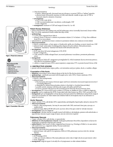
Figure 3. Patent duct arteriosus
Physical Exam for PDA
(in term infant)
• Heavy “machinery” murmur • High pulse rate
• Wide pulse pressure
• Hyperactive precordium
• Big bounding pulse
Figure 4. Coarctation of the aorta
• management:surgicalrepairifcriticallyillorifsymptomaticinolderinfants/children
