
Page 1250 - TNFlipTest
P. 1250
R4 Respirology
Approach to the Respiratory Patient Toronto Notes 2019 Pulmonary Function Tests
• usefulindifferentiatingthepatternoflungdisease(obstructivevs.restrictive)
• assesslungvolumes,flowrates,anddiffusioncapacity
• note: normal values for FEV1 are approximately ±20% of the predicted values (for age, sex, and height);
ethnicity may affect predicted values
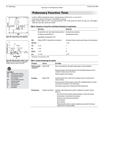
Table 5. Comparison of Lung Flow and Volume Parameters in Lung Disease
IC
FRC
FEV1
VC
TLC VT
1 SEC
ERV
Obstructive
Decreased flow rates (most marked during expiration) Air trapping (increased RV/TLC)
Hyperinflation (increased FRC, TLC)
DDx Asthma, COPD, CF, bronchiolitis, bronchiectasis* FEV1/FVC
TLC or N
RV or N
RV/TLC or N DLCO /or N
*Bronchiectasis can be obstructive or mixed
Table 6. Common Respirology Procedures
Restrictive
Decreased lung compliance Decreased lung volumes
ILD, pleural disease, neuromuscular disease, chest wall disease or N
N or N
RV
TIME
Figure 4A. Lung volumes and capacities
8 6 4 2 0
Obstructive
FEV1 VC
Normal Restrictive
Figure 4B. Expiratory flow volume curves
Adapted with permission from Elsevier. Weinberger SE. Principles of pulmonary medicine, 5th ed. 2008
Lung Volumes
Technique
Plethysmography (“body box”)
He Dilution
Bronchoscopy
Purpose
Measure FRC
Measure FRC
Diagnosis and therapy
Description
1 second time marker
86420
LUNG VOLUME (L)
ERV – FEF – FEV1 –
FRC – IC – RV – TLC – VC – VT –
ExpiratoryReserveVolume Forced Expiratory Flow Rate Forced Expiratory Volume (in one second)
Functional Residual Capacity Inspiratory Capacity Residual Volume
Total Lung Capacity
Vital Capacity Tidal Volume
After a normal expiration the patient inhales against a closed mouthpiece
Resultant changes in the volume and pressure of the plethysmograph are used to calculate the volume of gas in the thorax
Useful for patients with air trapping
A patient breathes from a closed circuit containing a known concentration and volume of helium
Since the amount of helium remains constant, FRC is determined based on the final concentration of the helium in the closed system
Only includes airspaces that communicate with the bronchial tree
A flexible or rigid bronchoscope is used for visualization of a patient’s airways Allows for:
Bronchial and broncho-alveolar lavage (washings) for culture and cytology Endobronchial or transbronchial tissue biopsies
Removal of secretions/foreign bodies/blood
Laser resections
Airway stenting
Mediastinal lymph nodes can also be sampled using a special bronchoscope equipped with an U/S probe (EBUS)
FLOW RATE (L/sec)
VOLUME
